Table 3. Ill-defined borders |
||
We give here some examples which might clarify the issue. However, to judge the degree of the blurred part of the borders always requires the analysis of the video.
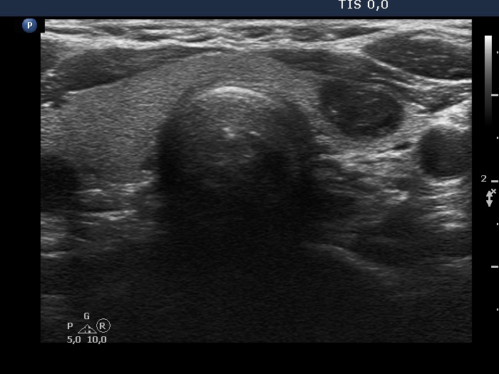
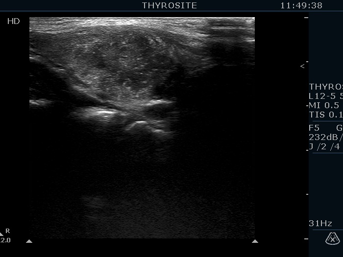
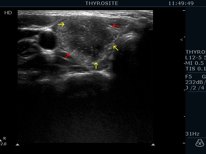
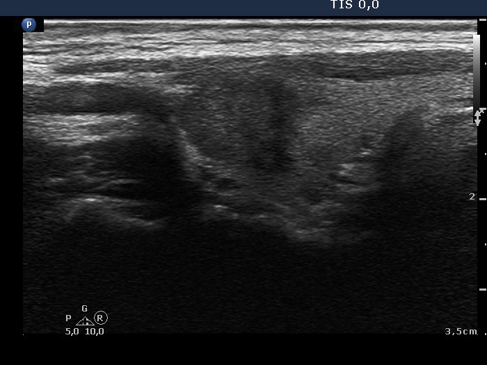
Benign nodular goiter (cytology) - case 2104 |
|
Transverse scan |
|
 |
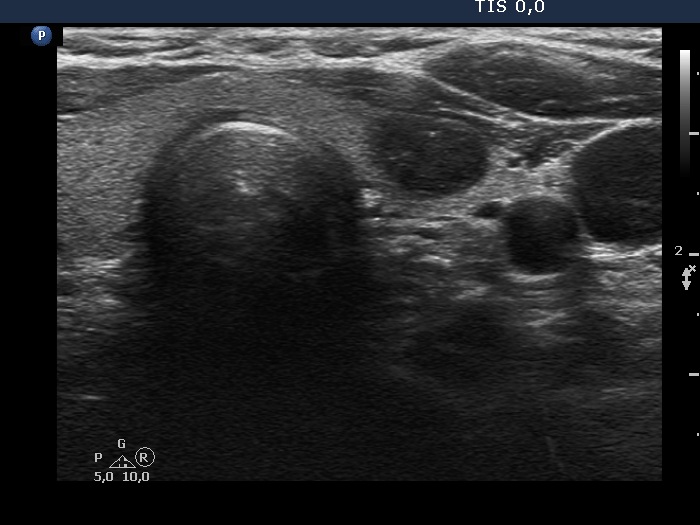 |
Longitudinal scan |
|
 |
 |
|
|
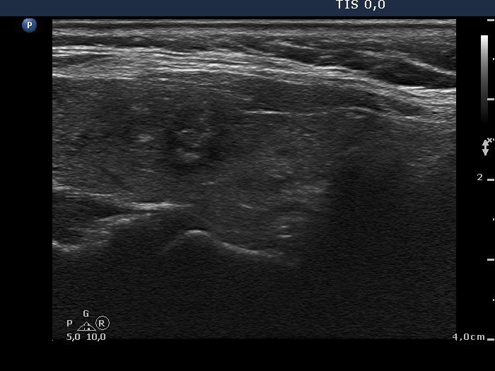
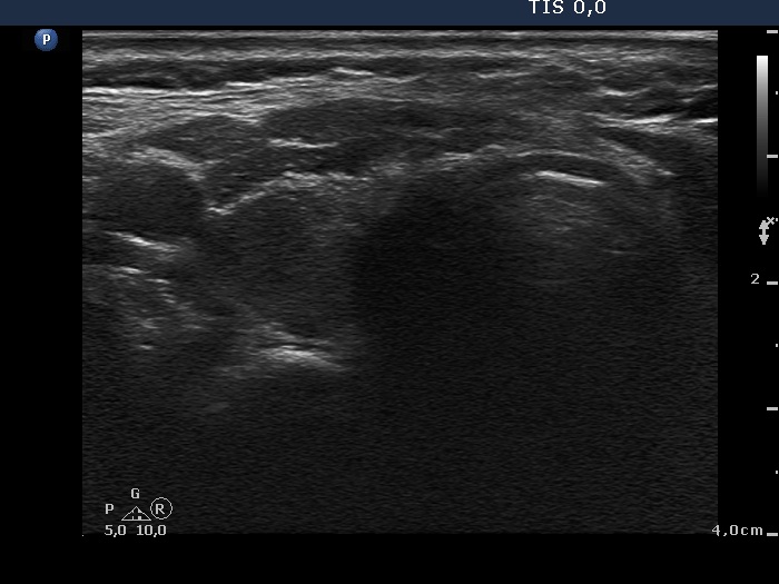
Papillary carcinoma (histology) - case conp003 |
|
Transverse scan |
Longitudinal scan |
 |
|
 |
 |
|
|
Papillary carcinoma (histology) - case 2082 |
|
Transverse scan |
Longitudinal scan |
 |
 |
 |
 |
|
|
Papillary carcinoma (histology) - case conp009 |
|
Transverse scan |
Longitudinal scan |
 |
 |
|
|
Benign lesion (cytology) - case 2069 |
|
Transverse scan |
Longitudinal scan |
 |
 |
 |
 |
|
|
Papillary carcinoma (histology) - case conp010 |
|
Transverse scan |
Longitudinal scan |
 |
 |
|
|
Papillary carcinoma (histology) - case conp051 |
|
Transverse scan |
Longitudinal scan |
 |
 |
|
|
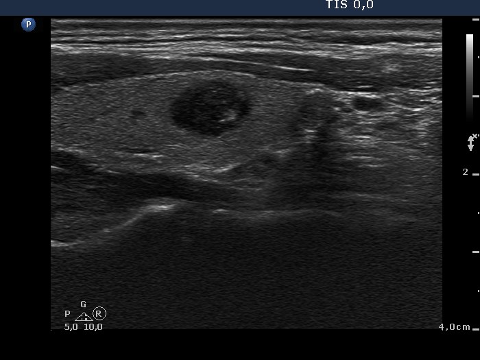
Benign lesion (cytology) - case 2142 |
|
Transverse scans |
Longitudinal scans |
 |
 |
 |
 |
|
|
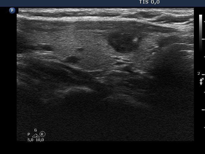
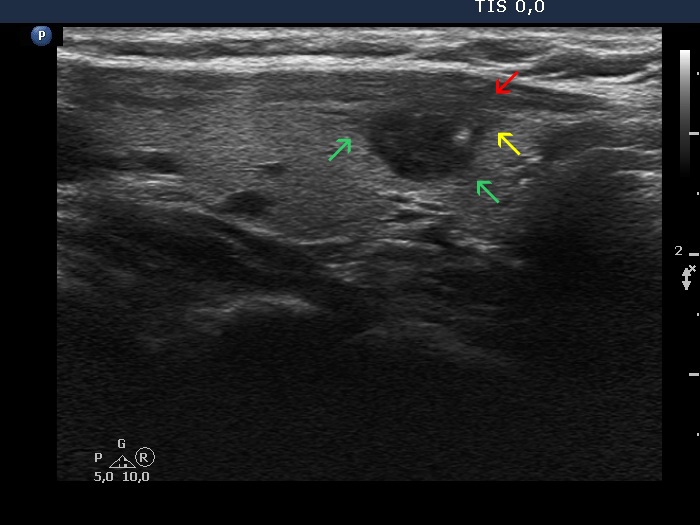
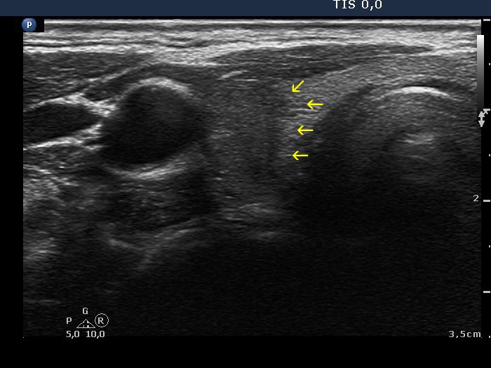
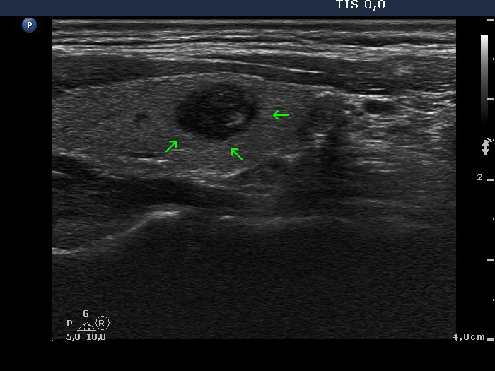
Papillary carcinoma (histology) - case conp020 |
|
Transverse scan |
Longitudinal scan |
 |
 |
 |
 |
The dorsal and medial borders of the nodule are partly ill-defined, partly lobulated. |
|
Papillary carcinoma (histology) - case conp032 |
|
Transverse scan |
Longitudinal scan |
 |
 |
|
|
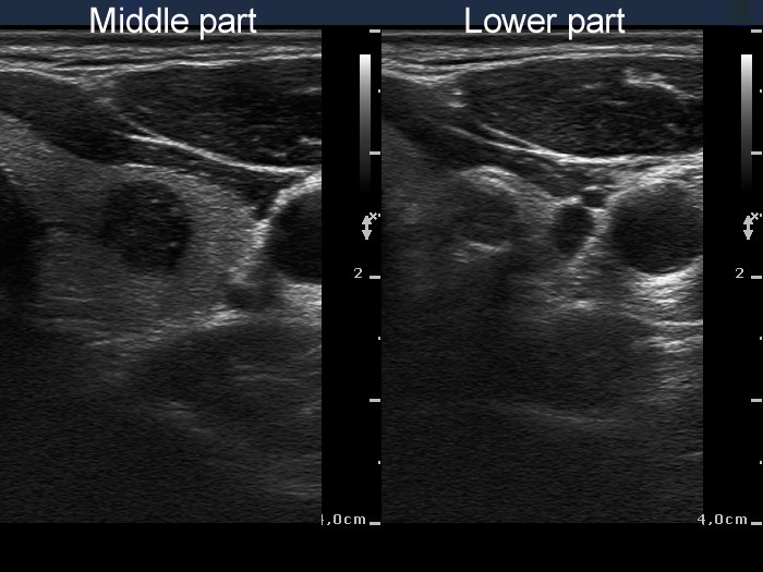
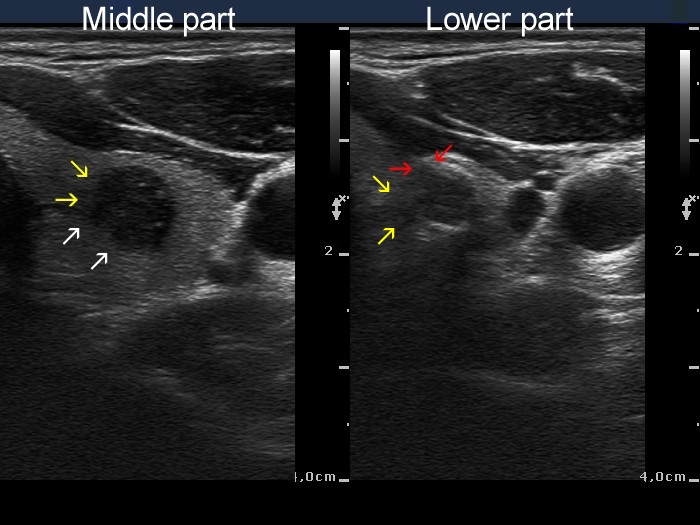
Papillary carcinoma (middle) and benign hyperplastic nodule (lower) (histology) - case conp034 |
|
Transverse scan |
Longitudinal scan |
 |
 |
 |
 |
|
|