Table 5. Spiculated margins |
||
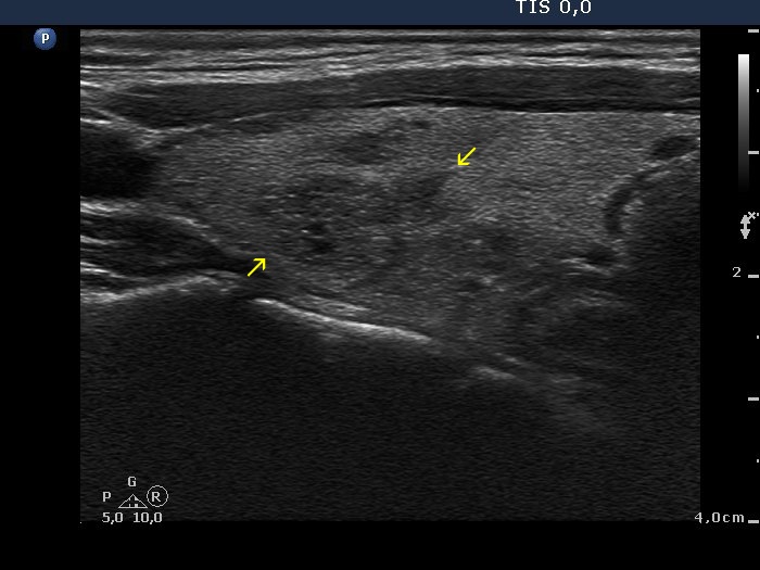
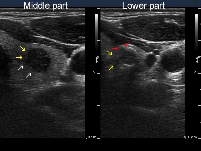
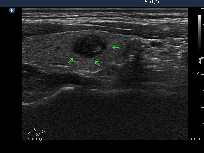
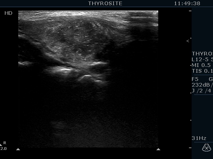
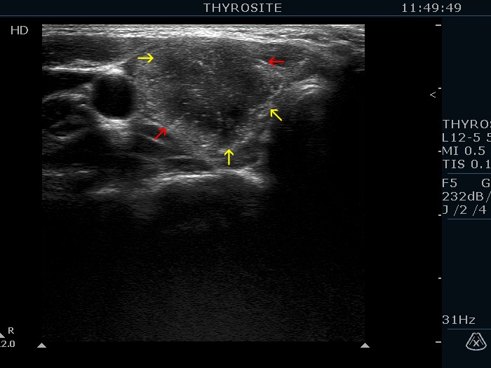
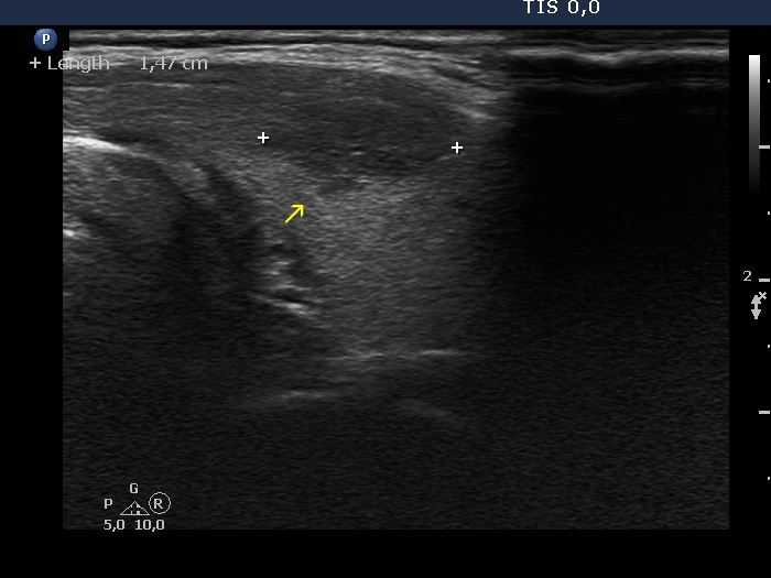
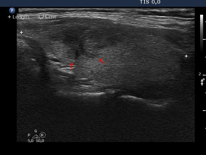
Spiculated margin belongs to the irregular borders which is encountered among suspicious signs of every TIRADS. This is characterized by the presence of projection having sharp angles. The distinction from lobulations is not always possible, but this has little if any relevance. Form a practical point-of-view, there are two issues. Firstly, there is no exact definition on which we can distinguish small irregularities on nodule surface without any clinical significance from pathognomonic spiculations. Secondly, one of the most typical presentations of Hashimoto's thyroiditis is the puzzle-like, spiculated borders of discrete hypoechogenic lesions.
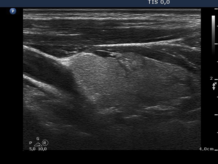
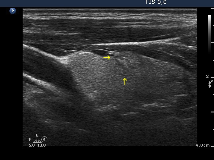
Papillary carcinoma (histology) - case conp003 |
|
Transverse scan |
Longitudinal scan |
 |
 |
 |
 |
|
|
Papillary carcinoma (histology) - case conp006 |
|
Transverse scan |
Longitudinal scan |
 |
 |
 |
 |
|
|
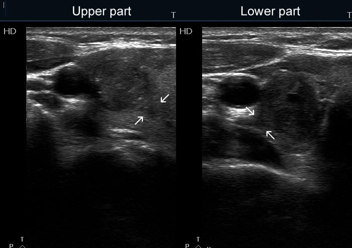
Papillary carcinoma (upper) and benign hyperplastic nodule (lower) (histology) - case conp027 |
|
Transverse scan |
Longitudinal scan |
 |
 |
 |
 |
|
|
Papillary carcinoma (upper) and benign hyperplastic nodule (lower) (histology) - case conp037 |
|
|
Longitudinal scan |
|
 |
 |
|
|
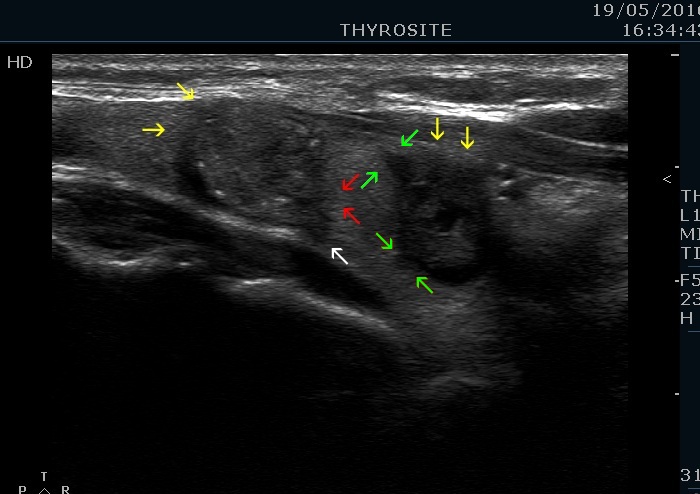
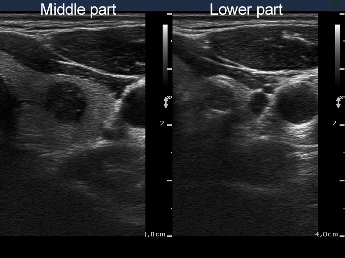
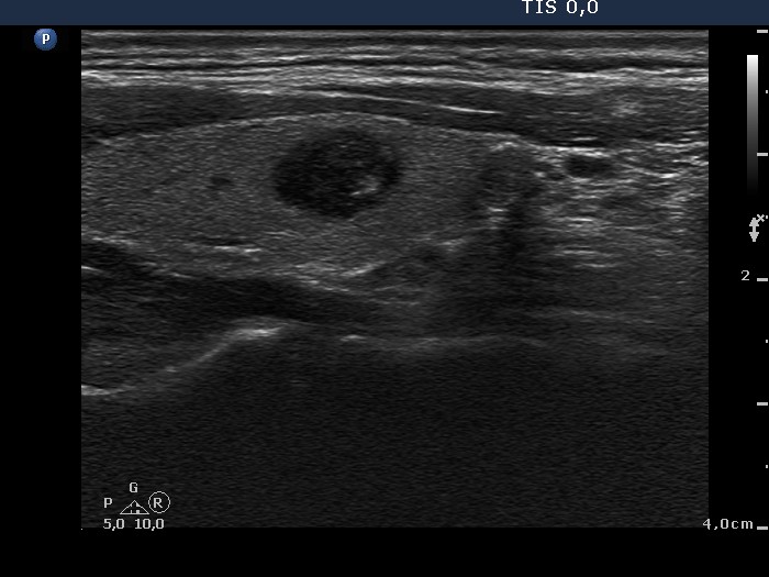
Papillary carcinoma (middle) and benign hyperplastic nodule (lower) (histology) - case conp034 |
|
Transverse scan |
Longitudinal scan |
 |
 |
 |
 |
|
|
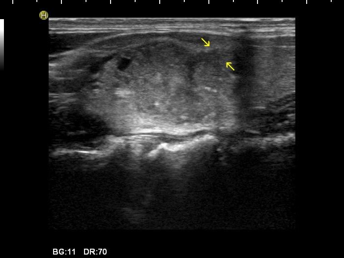
Papillary carcinoma (histology) - case 2082 |
|
Transverse scan |
Longitudinal scan |
 |
 |
 |
 |
|
|
Papillary carcinoma (histology) - case conp040 |
|
|
Longitudinal scans |
|
 |
 |
|
|
Oxyphilic variant of papillary carcinoma in an autonomously functioning nodule (histology) - case 2022 |
|
Transverse scans |
Longitudinal scans |
 |
 |
 |
 |
|
|