
|
|
Subacute granulomatous thyroiditis - case 1348
|
|


Initial examination (first row of images):
Clinical presentation: A 37-year-old woman requested checking of her thyroid status before a planned pregnancy.
Palpation: no abnormality.
Laboratory examination: TSH 2.06 mIU/L, aTPO 2 U/mL.
Ultrasonography: Both lobes were echonormal and intact. The vascularity was average.
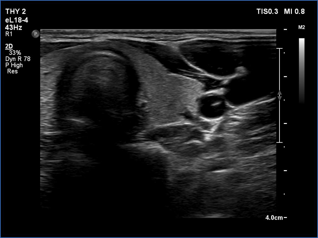
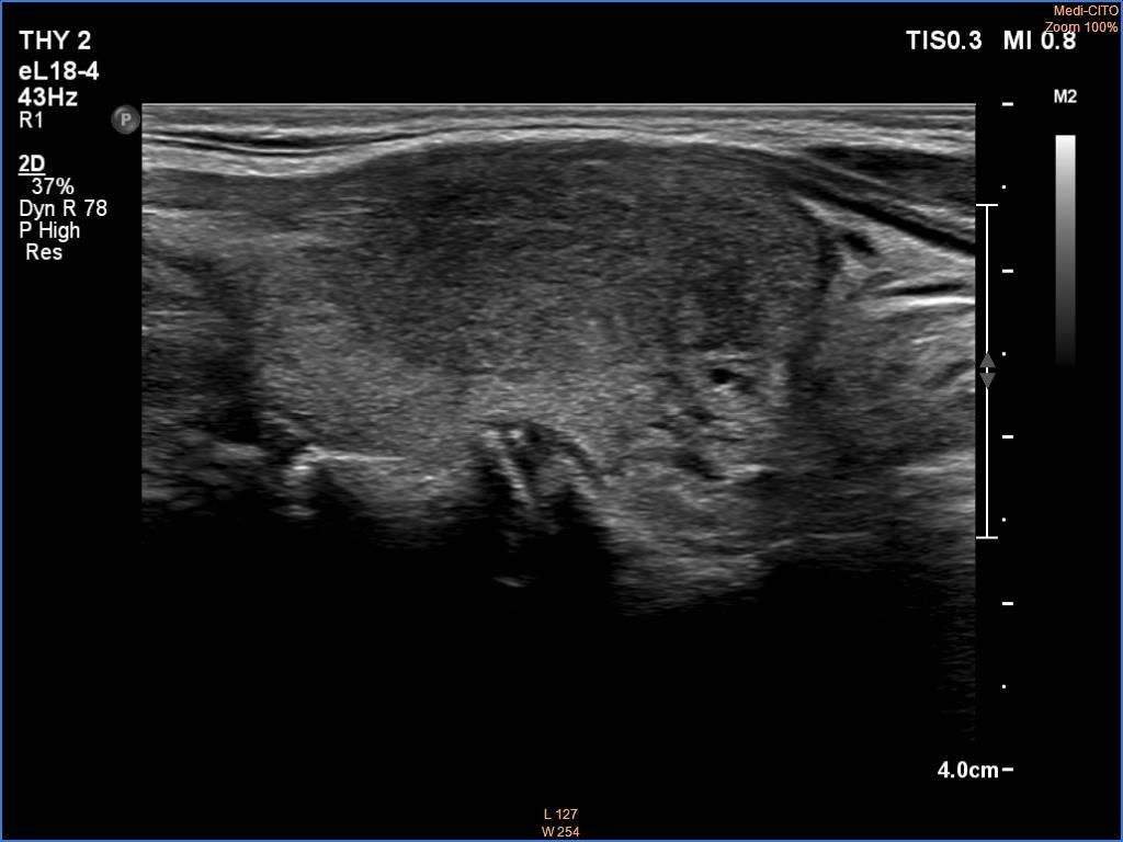
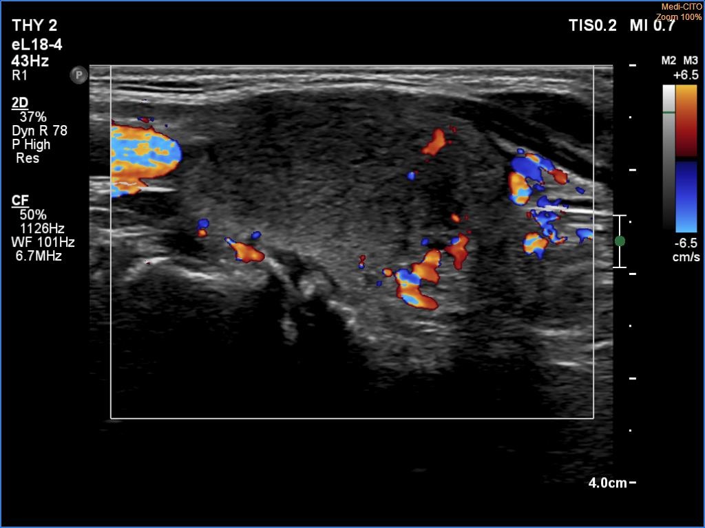
Examination 13 months later (second row of images):
Clinical presentation: The patient noticed neck pain and fever 6 weeks ago. Two weeks before the present visit, both sides of the neck became tender. Non-steroid anti-inflammatory drugs only temporarily ceased her complaints, two courses of antibiotics were ineffective.
Palpation: Both lobes were hard. The left lobe was painful, the right was very tender.
Laboratory tests: TSH 0.001 mIU/L, FT4 22,8 pM/L, FT3 7,03 pM/L, CRP 79 mg/L.
Ultrasonography: Both lobes has significantly increased in size and great part of the thyroid became hypoechoic. According to the hypoechoic areas, the vascularity was absent or only minimal.
Suggestion. Six-week steroid therapy.
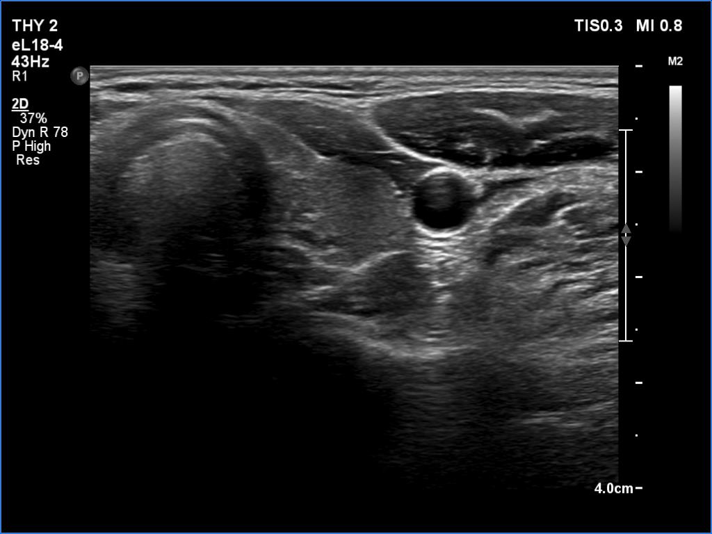
Examination a year after the first visit (third row of images):
Clinical presentation: The patient became free of complaints 6 hours after the first 32 mg-dose of methyl-prednisone and remained so for 4 weeks. When se decreased the dose to 8 mg every second day, the left thyroid became painful. Based on a telephone discussion, the steroid dose was increased. The complaints practically disappeared immediately.
Palpation: no abnormality.
Laboratory tests: TSH 4.01 mIU/L, FT4 9.7 pM/L, CRP 2.4 mg/L.
Ultrasonography: The ultrasound pattern, including the vascularity has normalized. The exception was the lateral part of the left lobe which remained hypoechoic and avascular.
Suggestion. Continue with the steroid therapy with decreasing those for another 3 weeks.