
|
|
Subacute granulomatous thyroiditis - case 795
|
|


First examination (1st and 2nd rows of images):
Clinical data: A 54-year-old woman presented with fever and a painful, suddenly enlarged goiter. The clinical picture was unaffected by two courses of antibiotic therapy. Her GP referred her for an evaluation with the suspicion of malignancy. The ESR was 87 mm/H, the CRP was 28.4 ng/L.
Palpation: a hard, uneven, and painful right thyroid lobe.
Functional state: moderate degree of hyperthyroidism with TSH 0.01 mIU/L, FT4 38.9 pM/L.
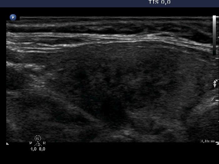
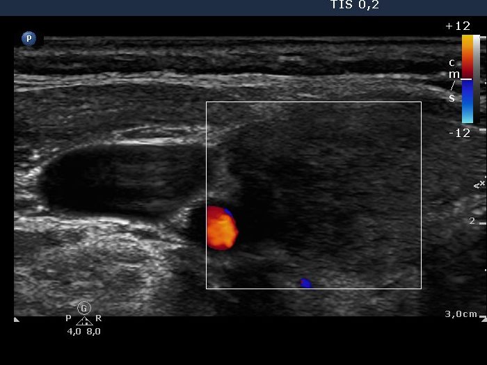
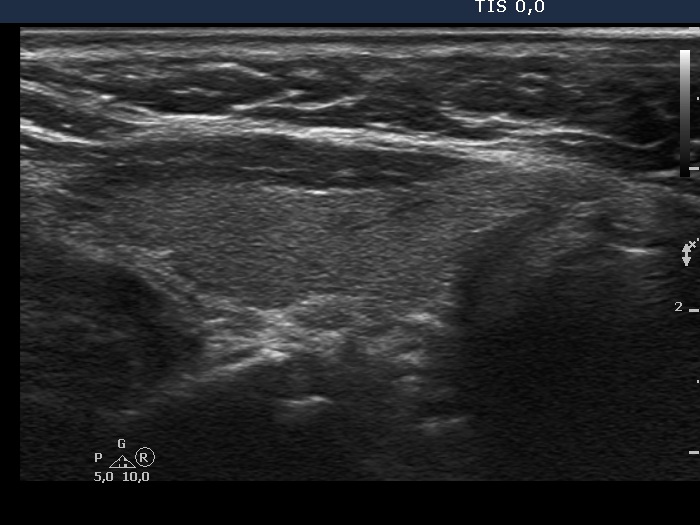
Ultrasonography revealed a moderately enlarged thyroid with a patchy hypoechogenic pattern in the right lobe and an intact left thyroid. There was no vascularization in the right lobe.
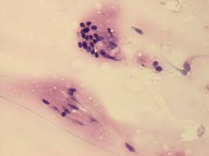
Cytological diagnosis: subacute, granulomatous thyroiditis.
Suggestion: a 6-week steroid therapy.
Follow-up examination 6 months later (3rd row of images):
Clinical data: The patient required one 6-week course of steroid therapy. Thereafter, she had no complaints except for fatigue lasting for 4 months.
Palpation: no abnormality.
Functional state: euthyroidism with TSH 2.94 mIU/L, FT4 11.2 pM/L. The CRP was 1.1 ng/L.
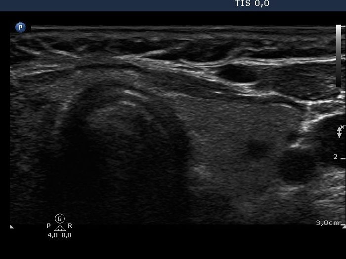
Ultrasonography: The hypoechogenic, ill-defined areas in the right lobe have disappeared, the size of the right has decreased. The vascularization was decreased but not absent.