Follicular adenoma - Case 20: a microfollicular
adenoma |
 |
 |
| 3-8 cells form a follicle which lacks
colloid. |
Follicular adenomas - Case 7:
a normofollicular adenoma |
 |
 |
| 8-20 cells form a follicle
which frequently contains colloid. |
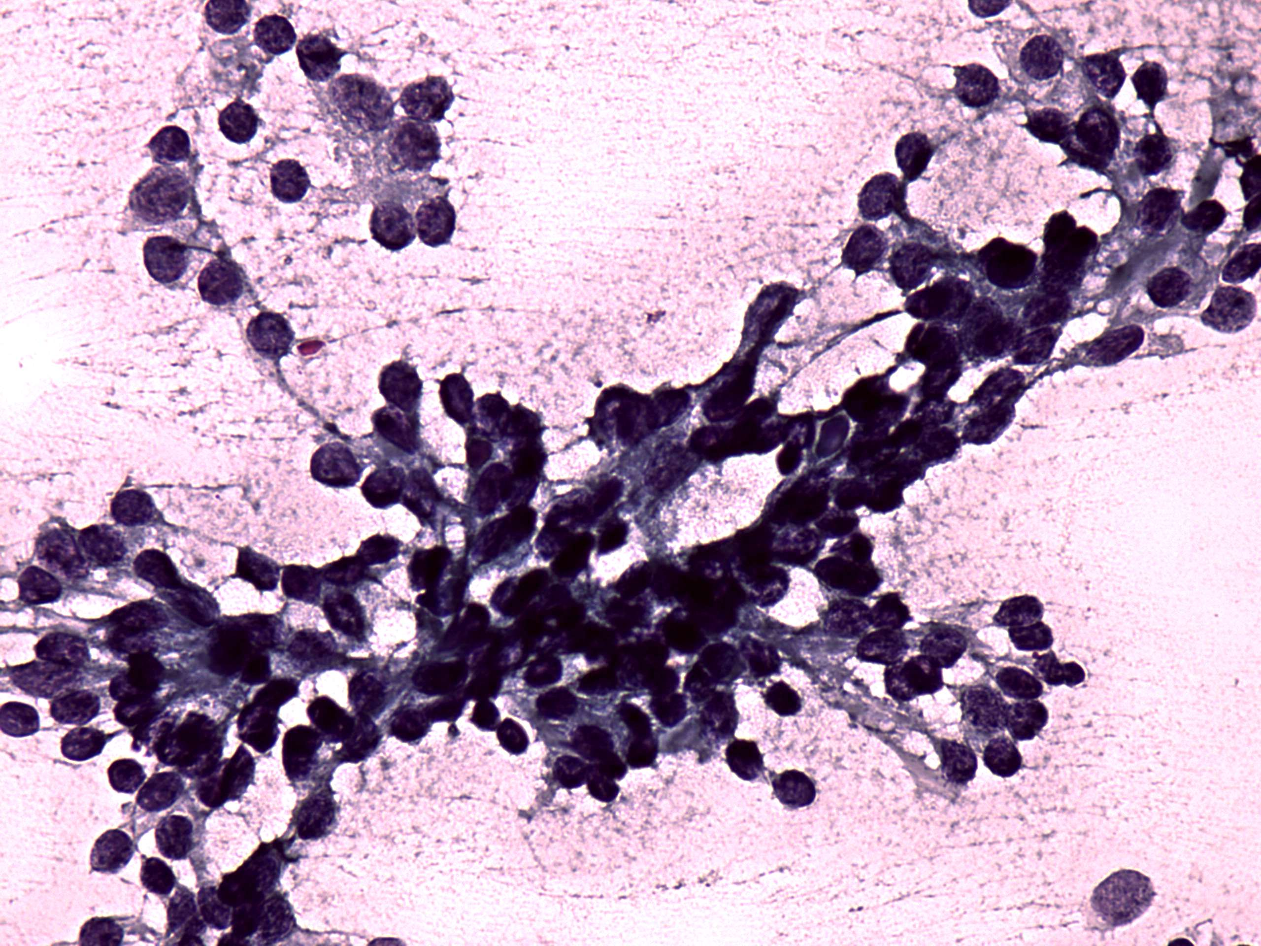
Follicular adenoma - Case 8:
a dominantly macrofollicular adenoma |
 |
 |
| More than 20 cells form a
follicle which contains colloid. Because of the size of the
follicle cystic degeneration is a frequent finding in such adenomas. |
|
Most adenomas contain all 3 types of follicles and the dominant subtype
decides whether an adenoma is microfollicular, normofollicular or
macrofollicular. It means that on a cytological sample we cannot decide
with enough safety from which type of adenoma we gained the smear.
Moreover, follicles are the basic structure of the thyroid, therefore
in every thyroid sample we can found follicles of one or more subtypes.
In the everyday practice microfollicular proliferation has relevance.
The basic rules are as follows:
- The greater the proportion of cells forming
folliculi, the greater the chance of a follicular tumor.
- The greater the proportion of well-preserved
microfolliculi, the greater the chance of microfollicular adenoma and
minimally invasive follicular carcinoma.
- The greater the proportion of small, irregular
folliculi, the greater the chance of an atypical follicular tumor.
|
Other rare tumors - Case 13:
a hyalinizing trabecular adenoma |
 |
 |
| The basic unit of a trabecular
adenoma is also a follicle but these are arranged in trabecular
cords. These are characteristically very cellular. |
|
|
 |
 |
Oxyphilic adenomas are a subgroup of follicular
adenomas,
therefore the basic structure in these tumors is also a follicle.
Because of abundant cytoplasm it is less conspicious the presence of
the follicular pattern in a cytological sample, dispersed cells
predominate most smears. This property and the presence of abundant
cytoplasm makes the pattern of an oxyphilic tumor similar to
medullary carcinoma.
|


