Follicular adenoma - Case 7 of a new approach |
Benign hyperplastic nodule - Case 7 |
 |
 |
 |
 |
 |
 |
 |
 |
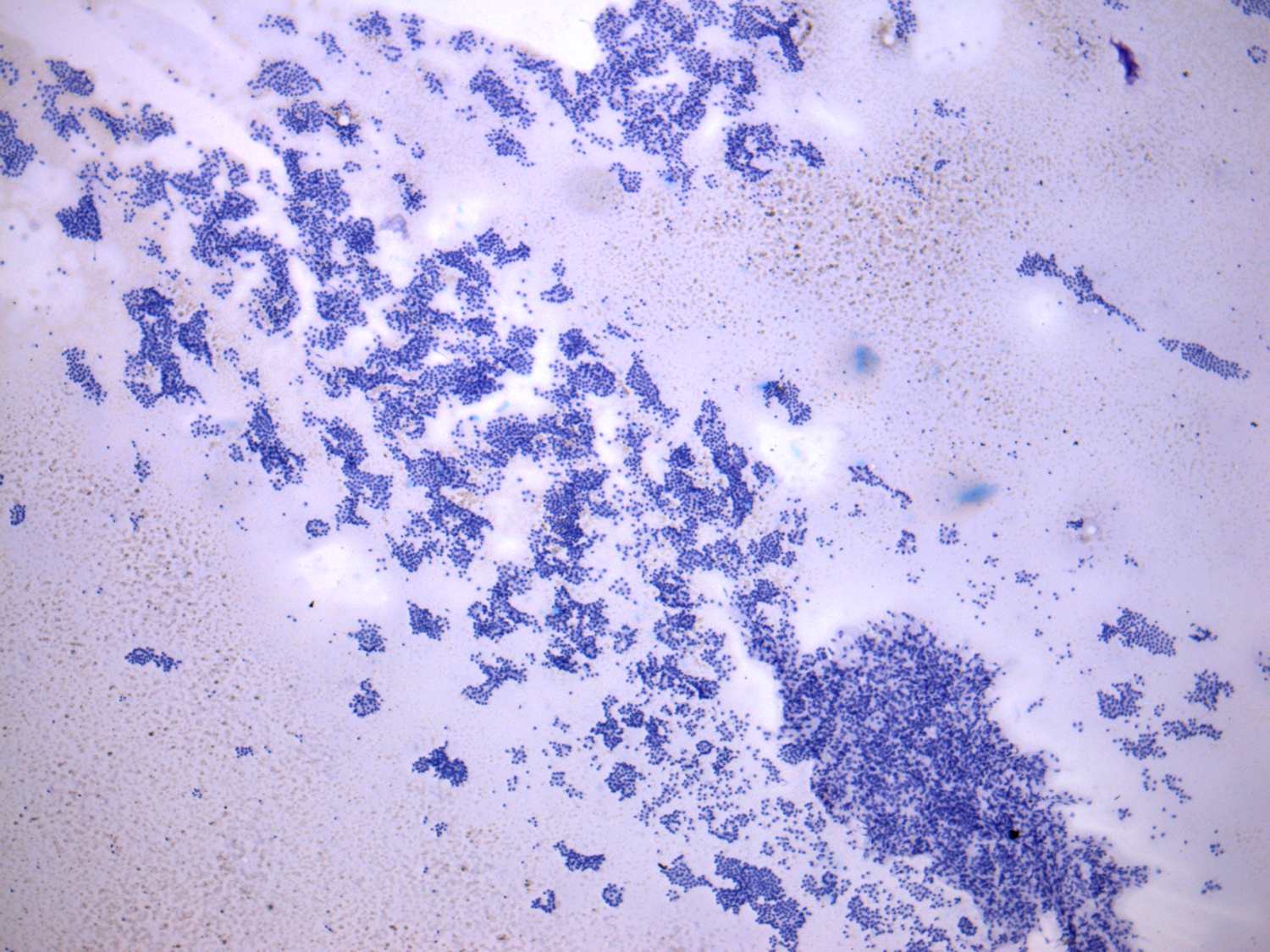
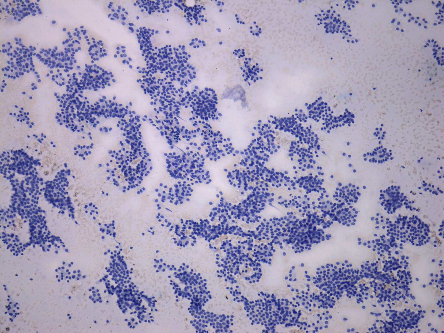
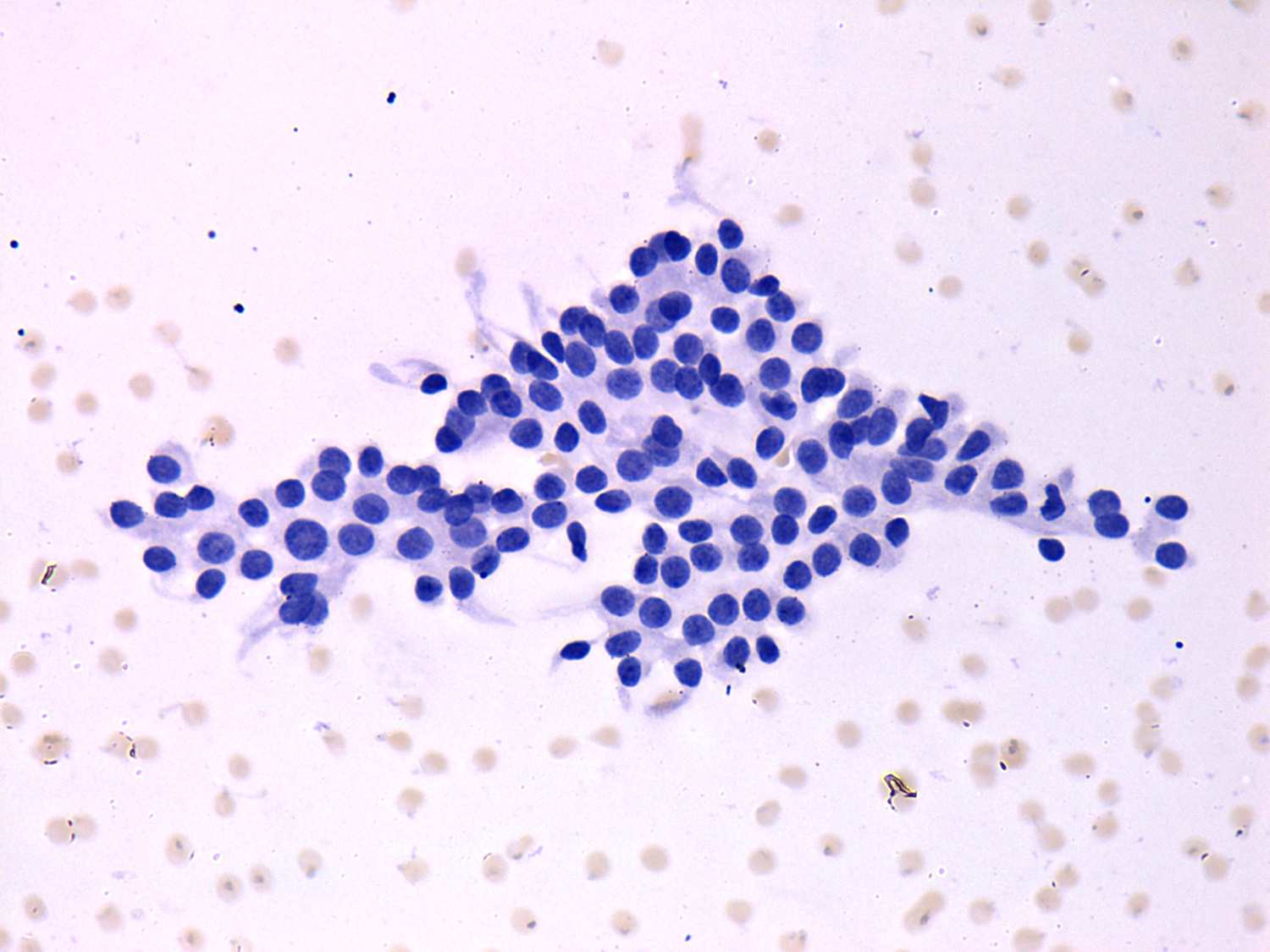
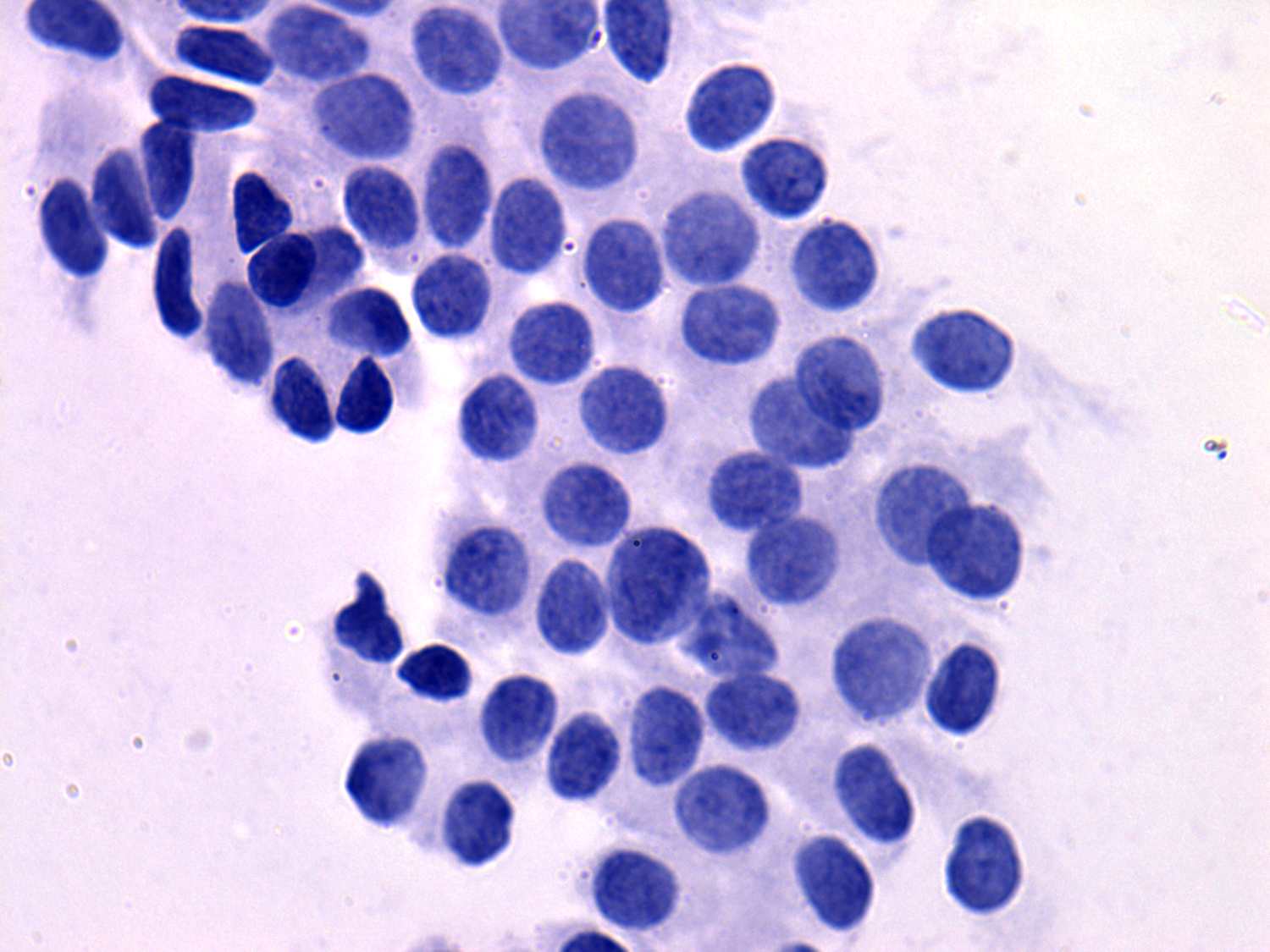
Microfollicular proliferation is demonstrated. Follicular cells are
uniform in shape and relatively uniform in size. Because of the lack of
prominent nucleoli beside a follicular tumor a hyperplastic nodule with
follicular proliferation have to be considered.
|
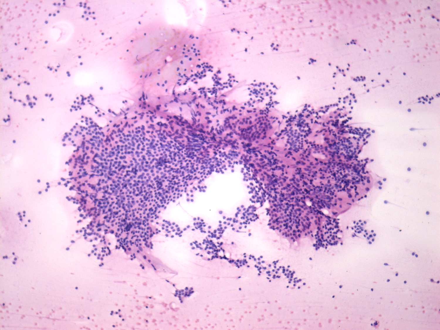
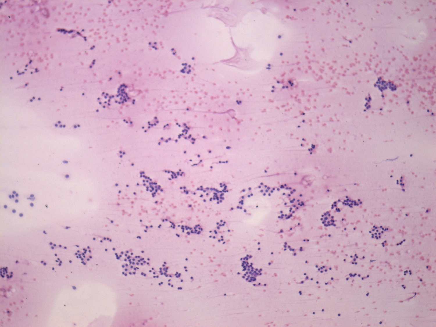
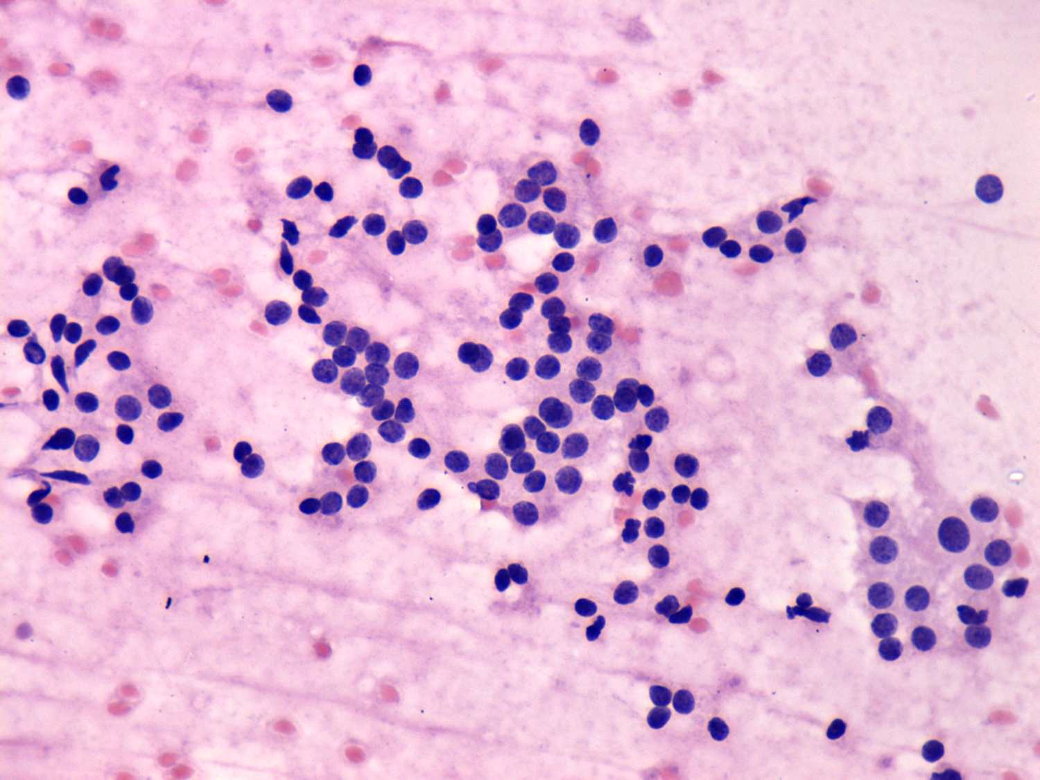
This pattern corresponds to a hyperplastic nodule with microfollicular
proliferation because of the presence of colloid and hyperplastic
papillary clusters. Nevertheless, the cytological pattern does not
exclude the possibility of follicular tumor.
|
 |
 |
 |
 |
 |
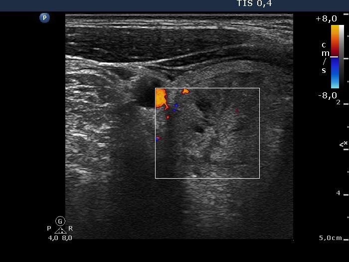 |
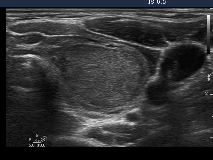
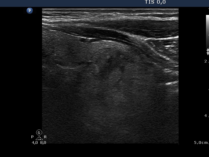
The nodule presents a halo sign and perinodular blood flow, therefore
this lesion is a follicular tumor with more than 90% probability.
|
Both the presence of a halo sign and perinodular blood flow are
doubtful.
In such cases the possibility of a hyperplastic nodule is greater than
follciular tumor.
|
|
Taking the cytological and sonographic
pattern into account, we gave a combined cytological-sonographic
diagnosis of a follicular tumor with less than the average risk of
carcinoma.
|
Taking the cytological and sonographic
pattern into account, we gave a combined cytological-sonographic
diagnosis of a benign follicular proliferation.
|
| |
|
| |
|

