Papillary carcinomas - Figure 19. Differential diagnostics: follicular tumor.
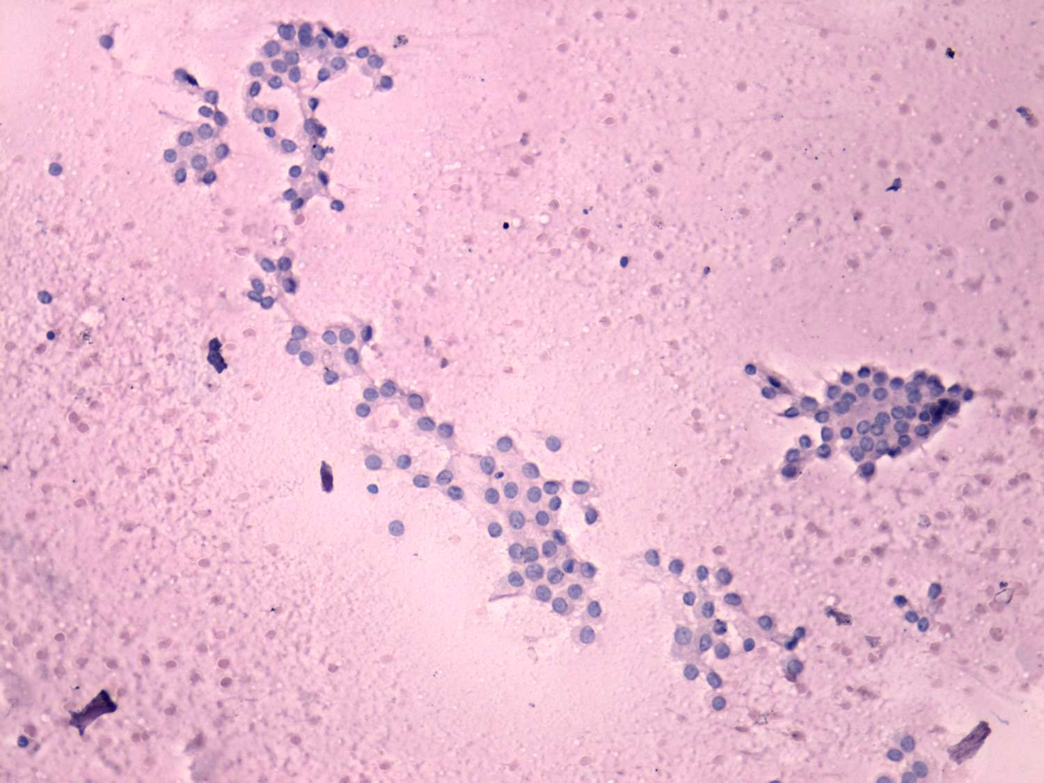
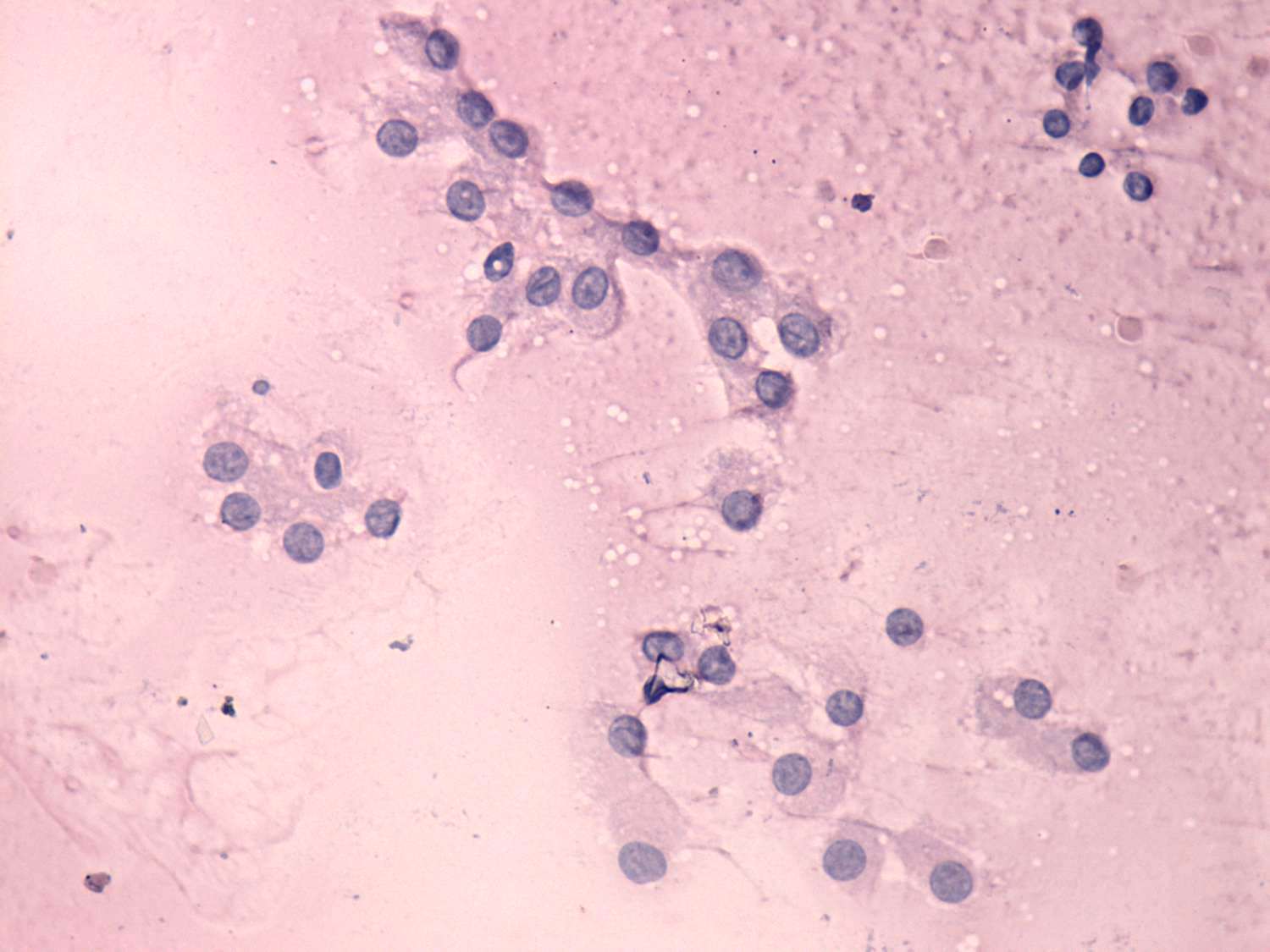
The presence of grooves and inclusions is a rare phenomenon in the case of a follicular tumor except for Hürthle-cell variant. In such cases the cytological differential diagnostics may be impossible. If we have any doubt the definitive cytological diagnosis of a papillary carcinoma should be avoided.
|
Follicular adenoma - case 30
|
Papillary
carcinoma - case 63
|
 |
 |
 |
 |
 |
 |
 |
 |
|
|
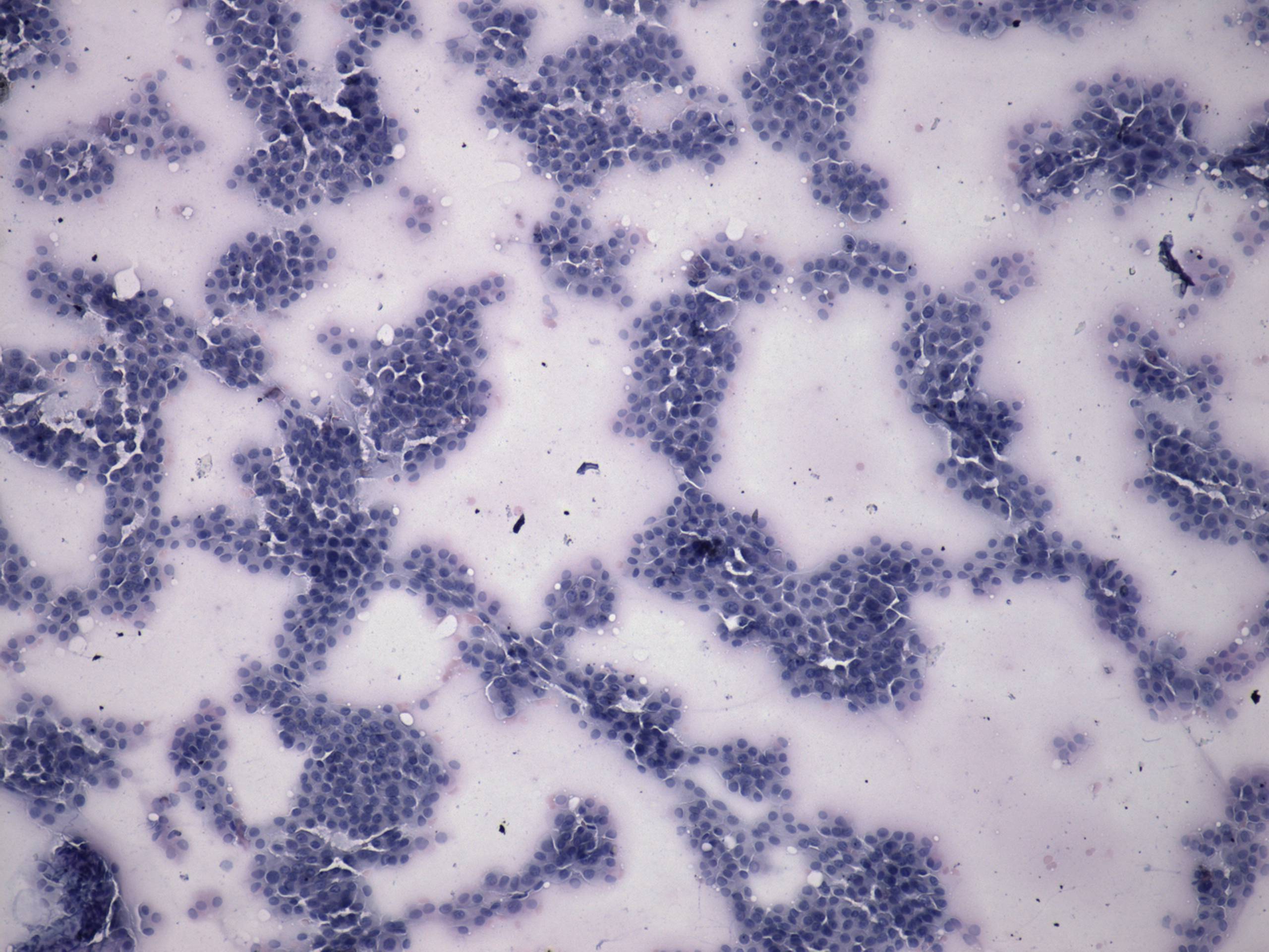
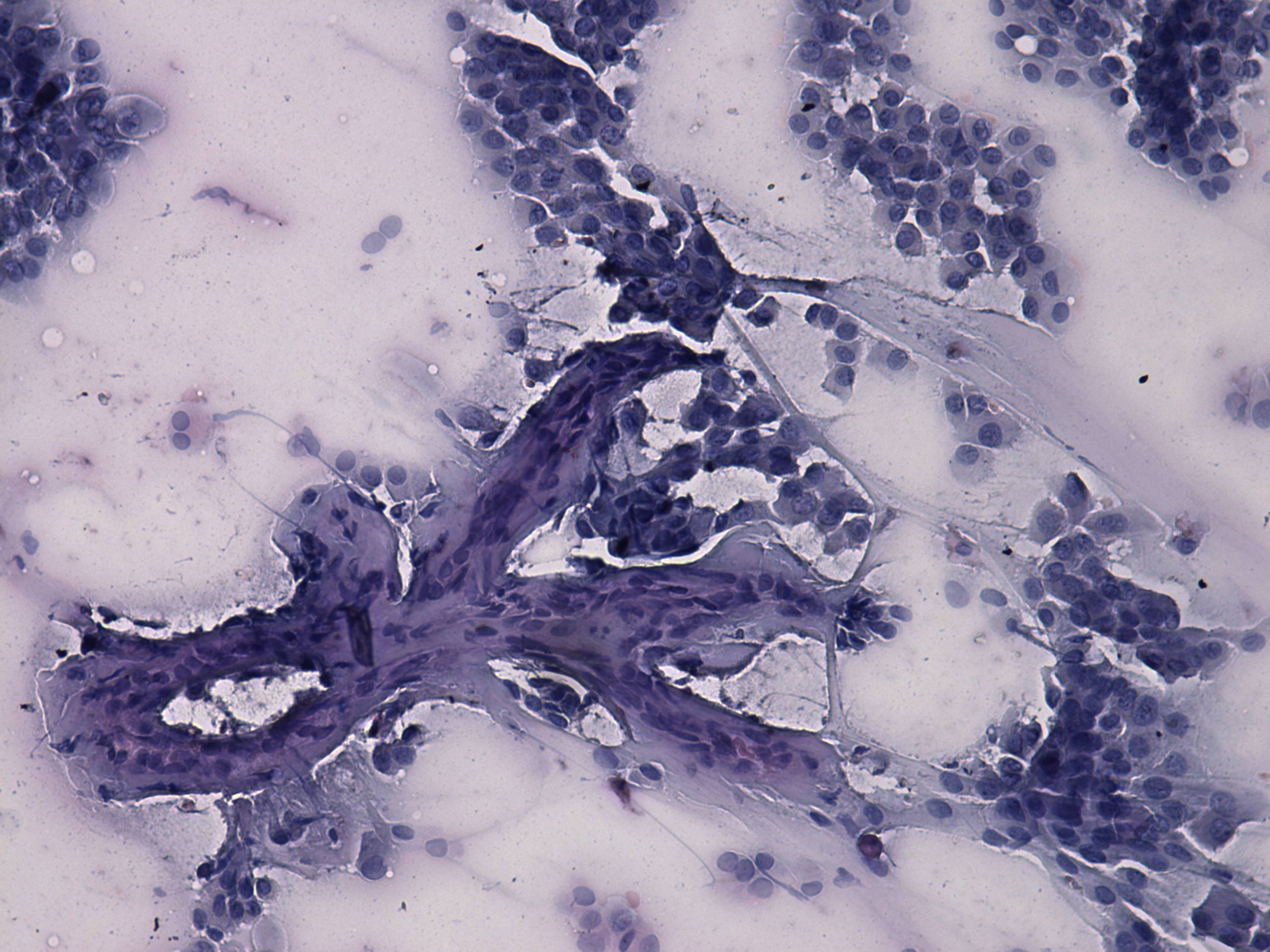
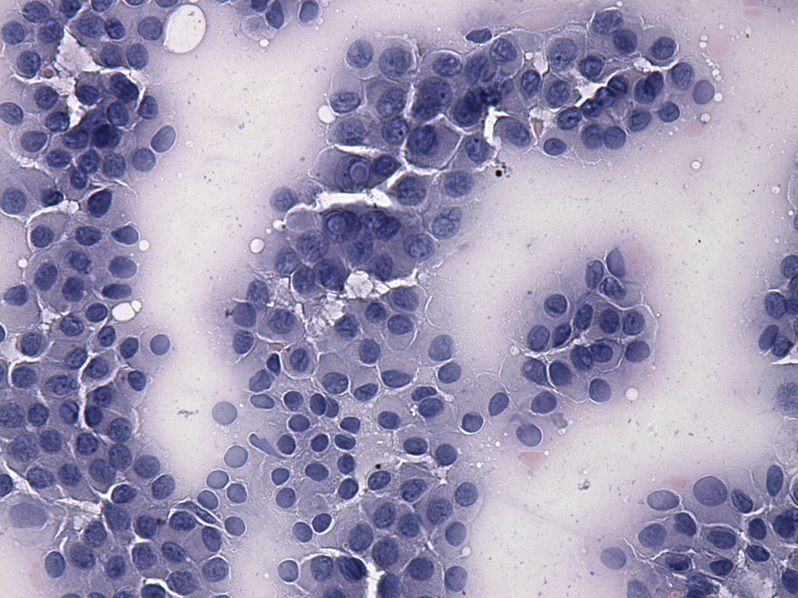
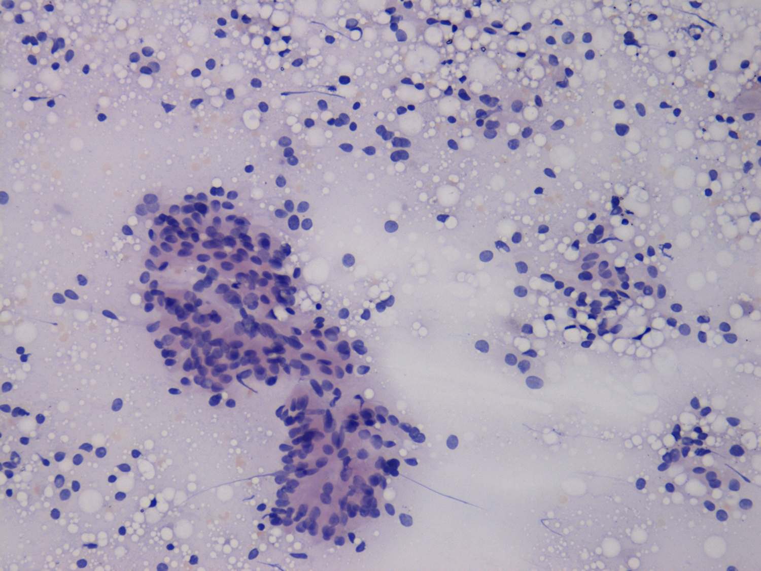
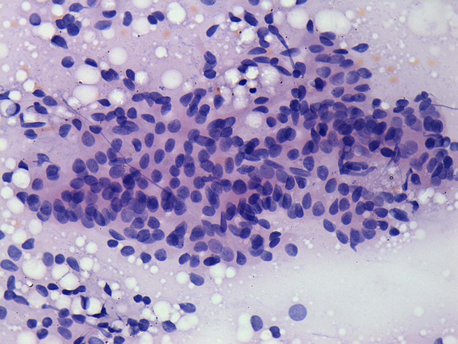
Follicular cells with abundant cytoplasm occur in monolayered sheets composed of microfollicules. Note the presence of fibrovascular core in the second image and the large number of inclusions. These signs have to raise the possibility of a papillary carcinoma. |
|
Follicular adenoma - case 30 |
Papillary carcinoma - case 63 |
 |
 |
 |
 |
 |
 |
 |
 |
|
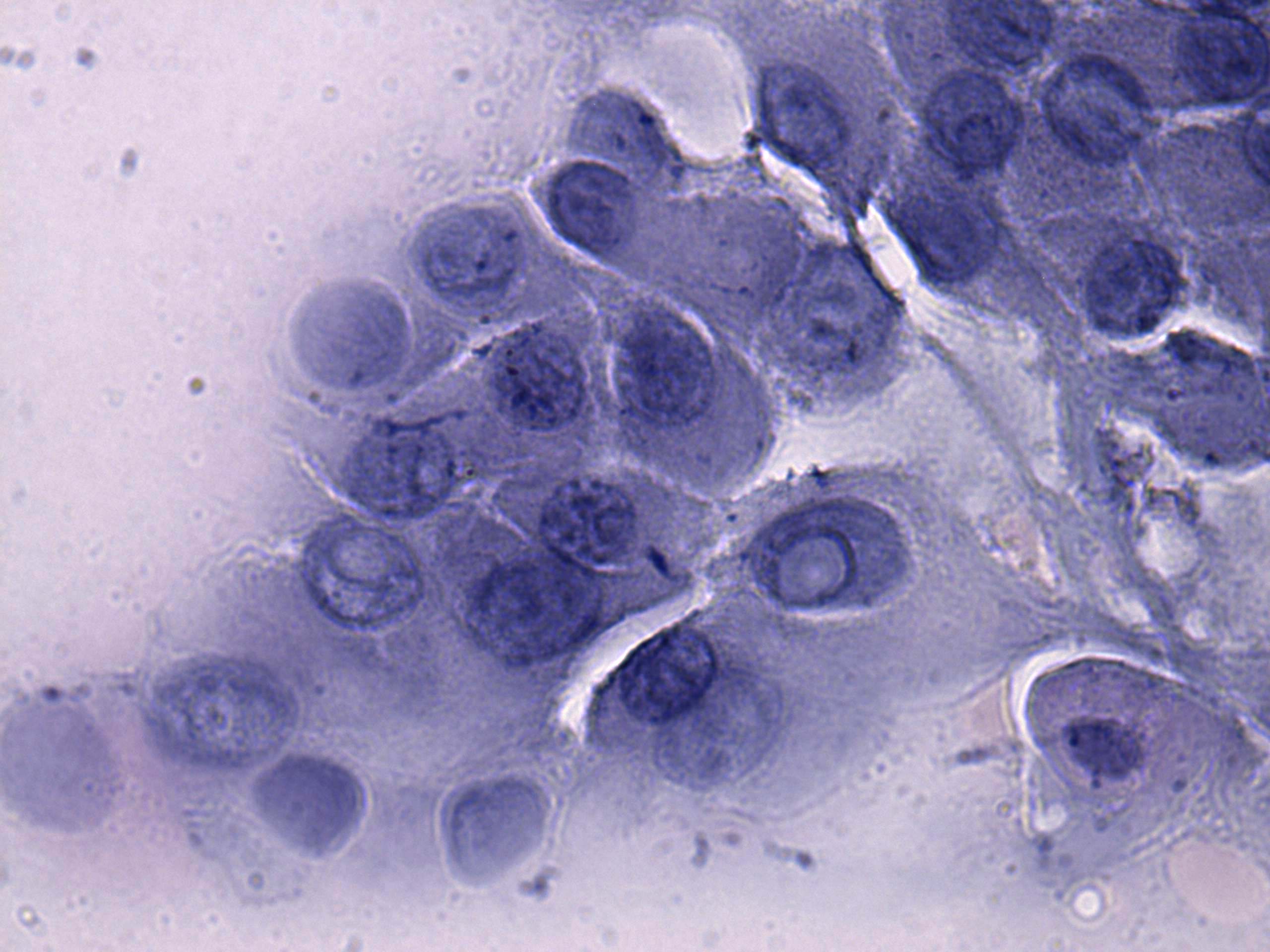
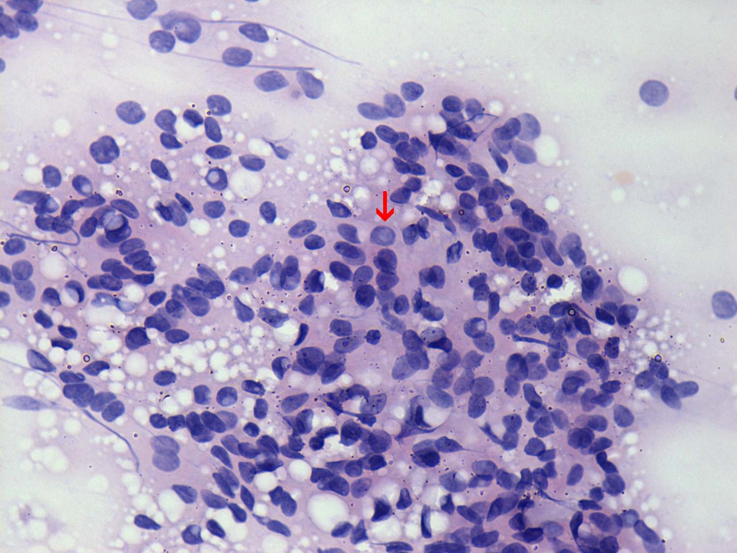
The nuclei are pale, one nucleus contains an inclusion while several others present groove. The presence of diffuse colloid in the background and the peripheral vacuoles at the edge of the papillary fragments demonstrated in the third image stands against but does not exclude the possibility of a carcinoma. |
The presence of colloid, the fragments seemingly typical of a hyperplastic nodule stand against the possiility of a carcinoma. Nevertheless, the nuclear crowding and a few inclusions other than projections of vacuoles to the nuclei (red arrow) are arguments against benign nature of the cells. Nevertheless, a definitive diagnosis of a papillary carcinoma is not possible.l |