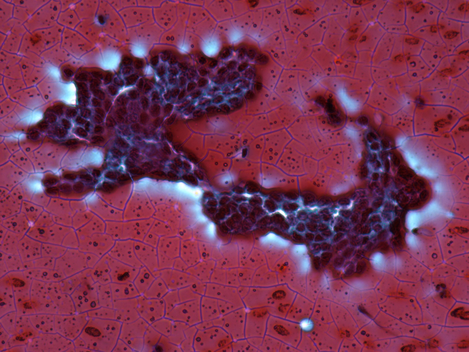
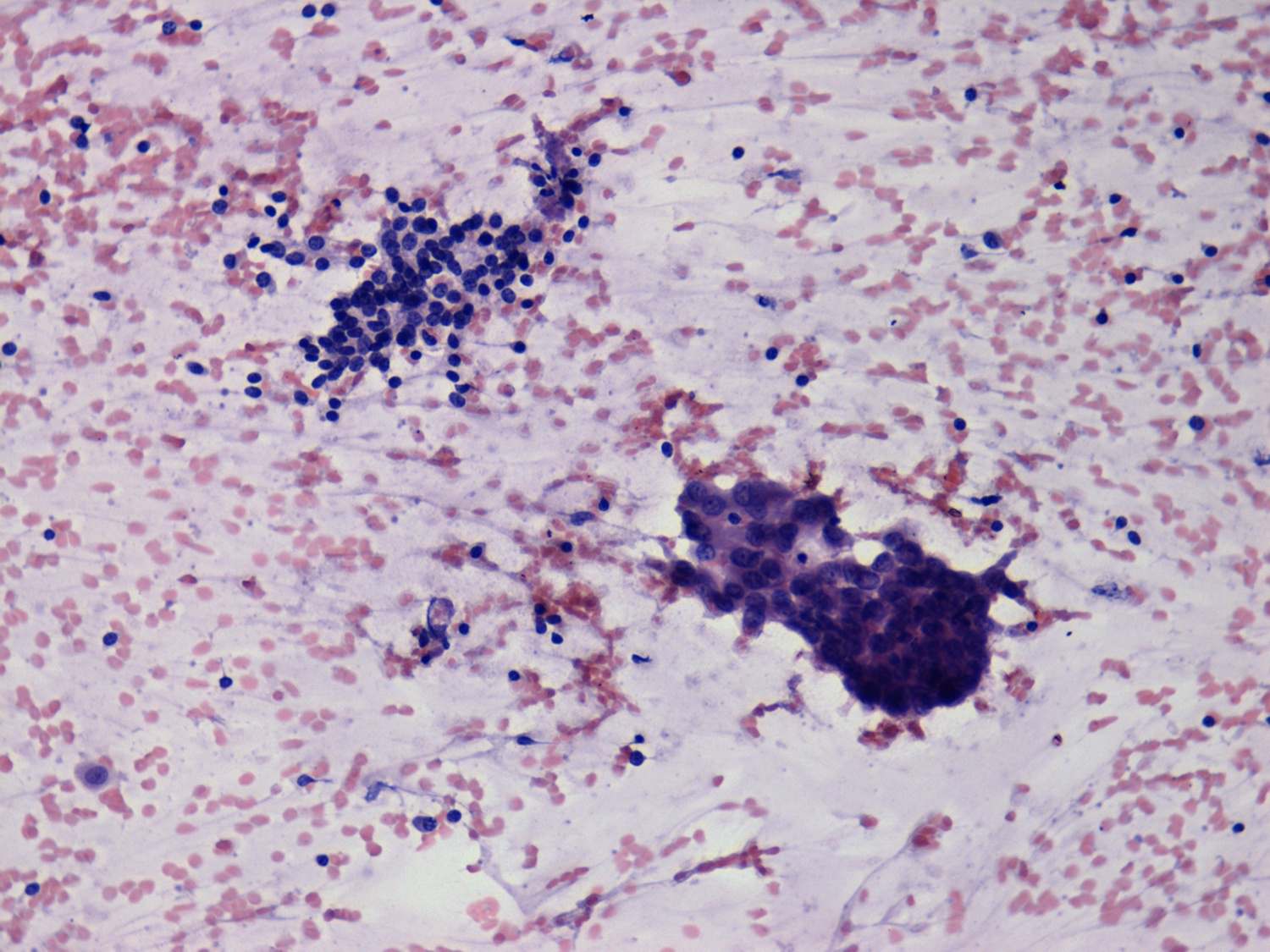
Papillary carcinomas - Figure . Cytological presentation - different population of cells on the same smear.
This type of atypia has little relevance in the everyday practice because most cases present typical signs of papillary cancer. If we had any doubt wash-out technique or thyroglobulin immuncytochemistry decides the issue.
Papillary carcinoma - case 18 |
|
 |
 |
| The left image presents typical signs of a colloid goiter, while the left image does an absolutely different pattern of the papillary carcinoma. | |
Papillary carcinoma - case 6 |
|
 |
 |
| Two different cell populations are demonstrated in a common field of vision. The upper cell group arose from a benign lesion while the lower cluster from the papillary carcinoma. | Left the benign, right the malignant cells at higher power field. |