Table 6. Differential diagnotics of lobulated and spiculated margins

doi: 10.24390/thyrosite.ctu.2.5.lect.06.2


As in many cases of differential diagnostic, the issue is whether an irregular border is caused by any forms of thyroiditis or a pathological nodule. The former is characterized by multiple while the latter with only one or two discrete lesions per thyroid lobe. A clear distinction simply on ultrasound pattern is not infrequently impossible.

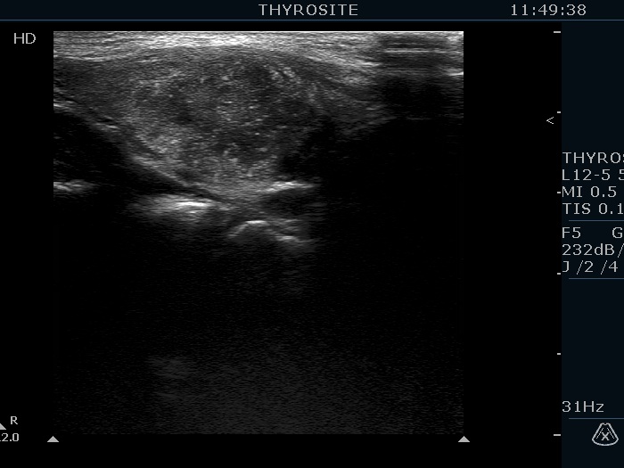
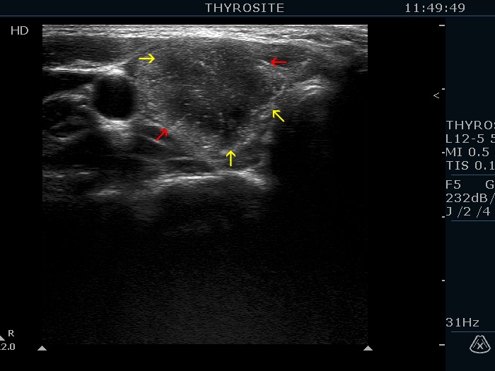
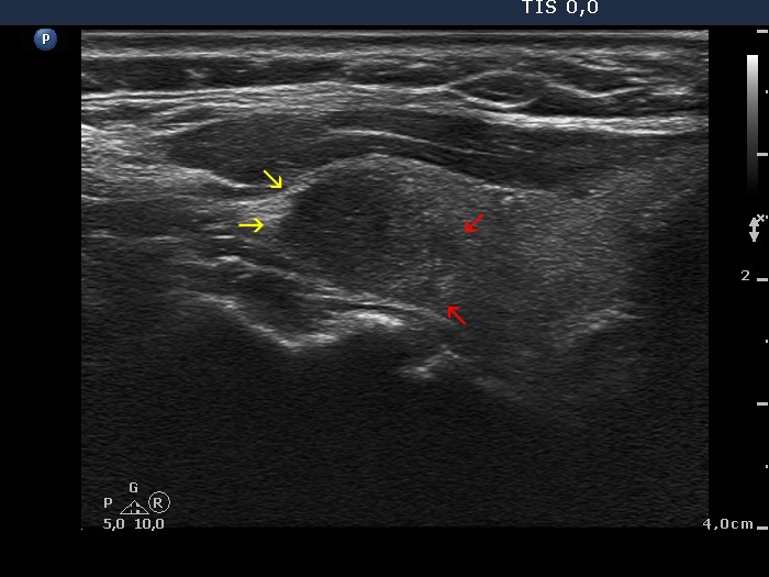
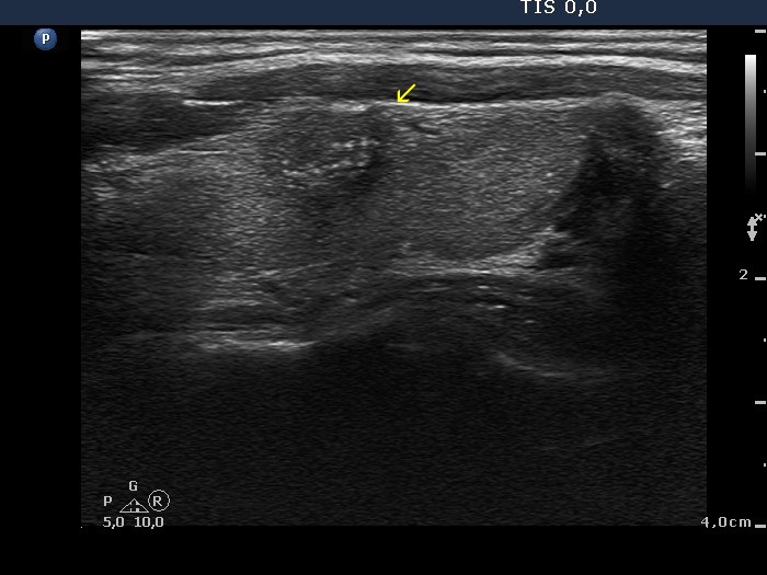
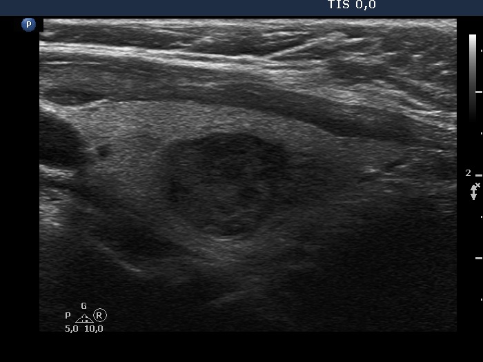
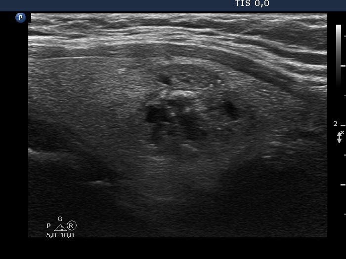
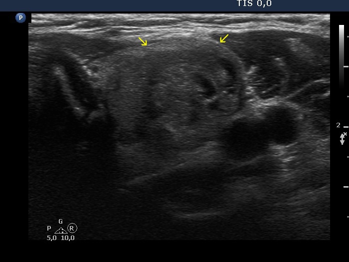
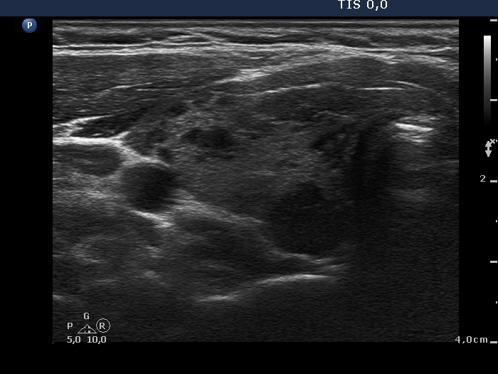
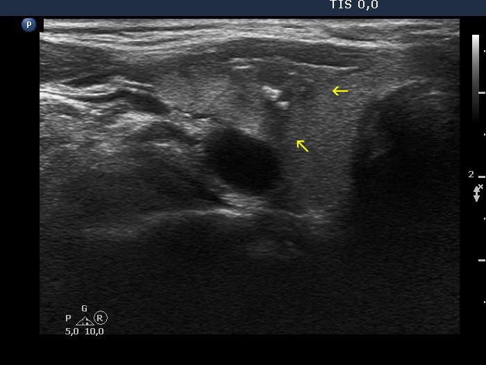
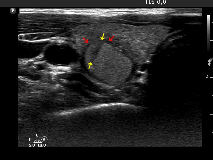
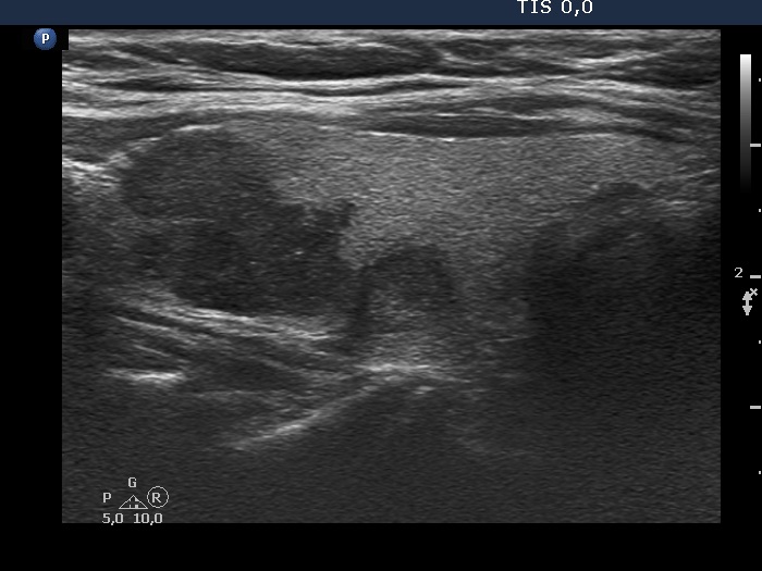
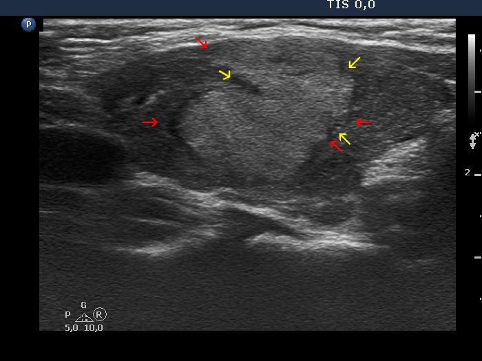
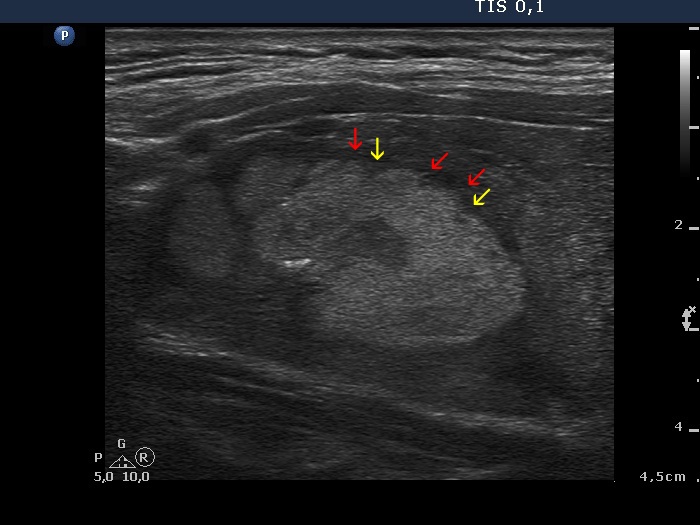
Papillary carcinoma (histology) - case conp003
Transverse scan
Longitudinal scan

The lesion has partly blurred and partly spiculated margins. Although this presentation corresponds to thyroiditis, the fact that there are one or two relatively large lesions in the lobe should raise the possibility of nodular origin, namely papillary carcinoma. (Three types of indistinct borders are presented in these images. Green arrows point to that part of the nodule which echogenicity is identical to the extrathyroidal strap muscle. The tumor has partly blurred (red arrows) and partly spiculated (yellow arrows) margins. The extent of blurred part of the border exceeds 50%.)


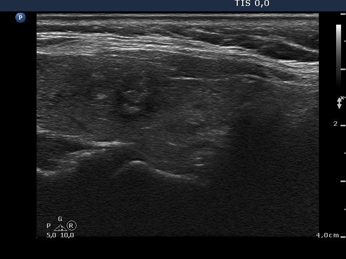
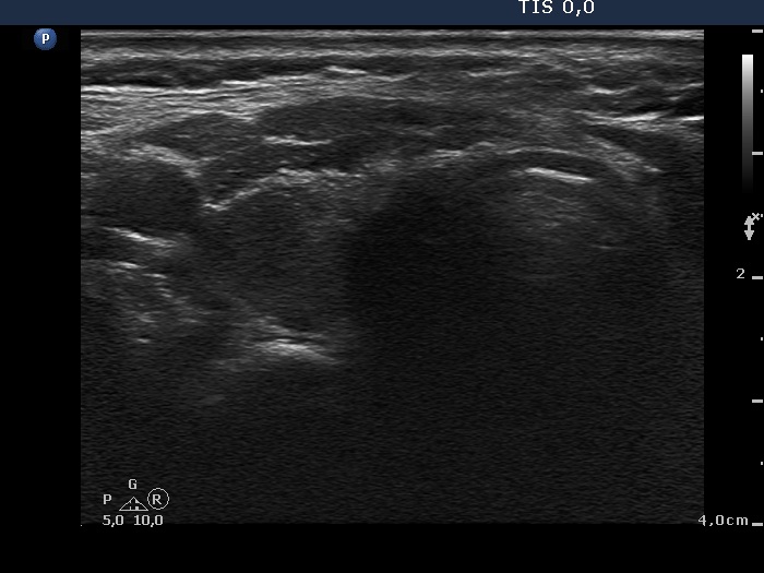
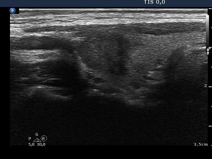
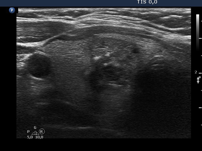
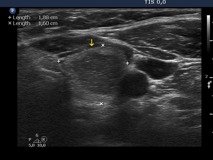
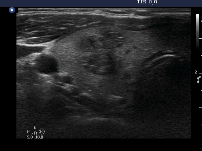
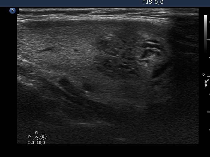
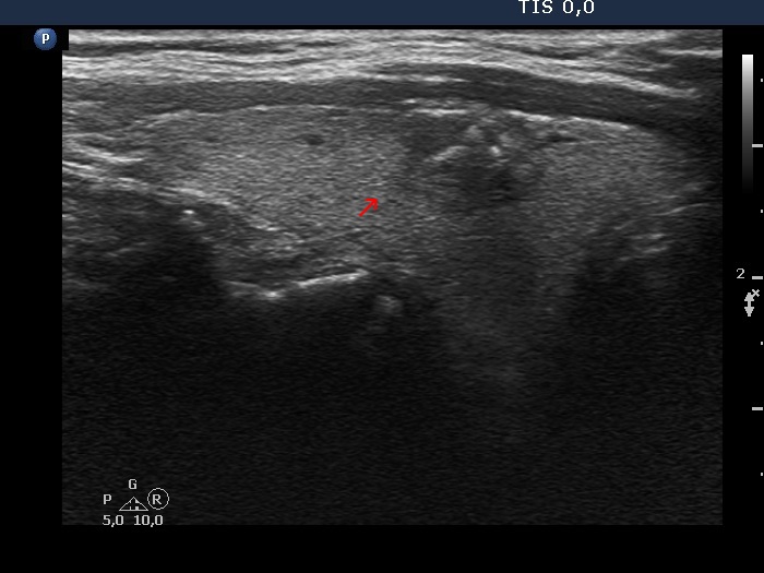
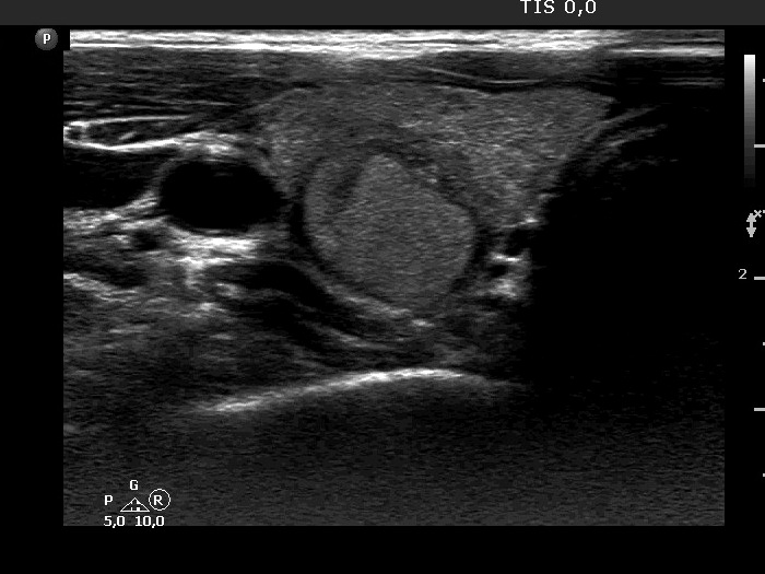
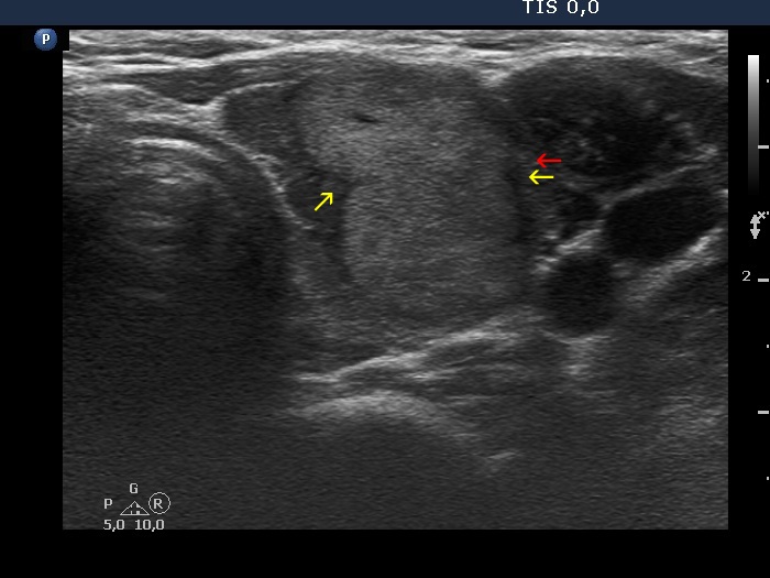
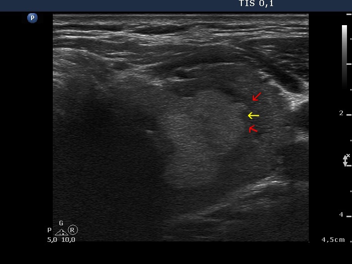
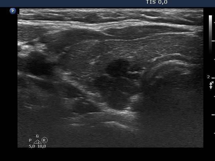
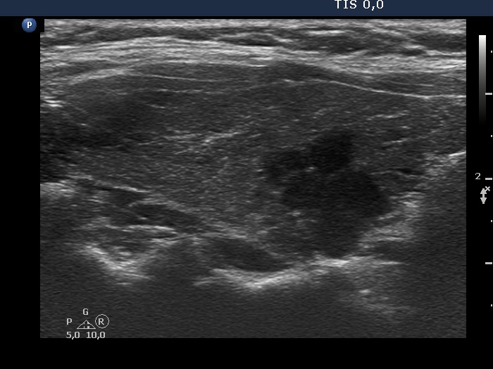
Papillary carcinoma (histology) - case 2082

Transverse scan
Longitudinal scan

In this case almost the entire border of the nodule is ill-defined, moreover the lesion has lobulated (yellow arrows) and spiculated (red arrows) margins, as well.

   
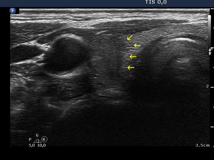
Papillary carcinoma (histology) - case conp009
Transverse scan
Longitudinal scan

In this case more than 50% of the nodule' borders are ill-defined and therefore it is difficult to judge the protrusions on the nodule' surface.


Benign lesion (cytology) - case 2069

Transverse scan
Longitudinal scan

The nodule has ill-defined borders in less than the third and more than the three-fourth of the circumference of the nodule, transverse scan and longitudinal view, respectively. (See the border between the red arrows.) The nodule also presents lobulation, the small irregularities marked with white arrows are not necessarily pathological, while those marked with yellow are abnormal findings.

   
Papillary carcinoma (histology) - case conp010
Transverse scan
Longitudinal scan

The borders of the nodule are both blurred and spiculated-lobulated.


Papillary carcinoma (histology) - case conp051
Transverse scan
Longitudinal scan

This is a typical example of ill-defined borders. Moreover, the nodule presents protrusions which are suspicious (red) and diagnostic (yellow) for lobulated margins. The blur of the margins hinders the judgement of protrusions.

   
Papillary carcinoma (histology) - case conp020
Transverse scan
Longitudinal scan
The dorsal and medial border of the nodule is partly ill-defined partly lobulated.
   

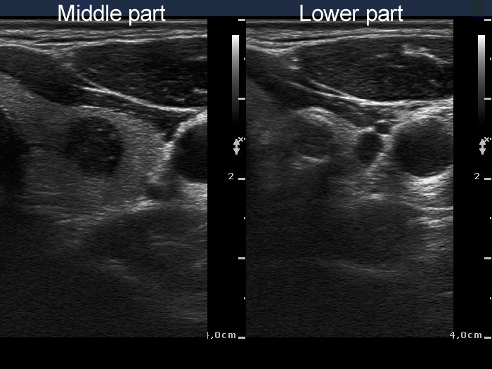
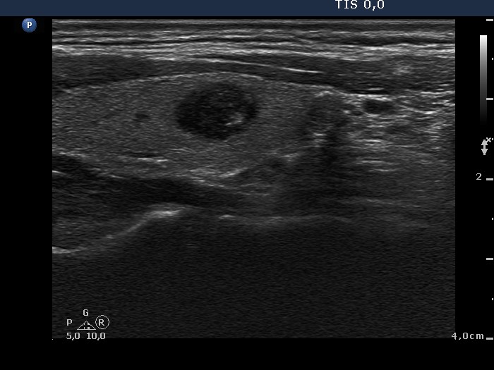
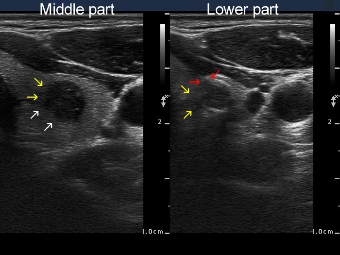
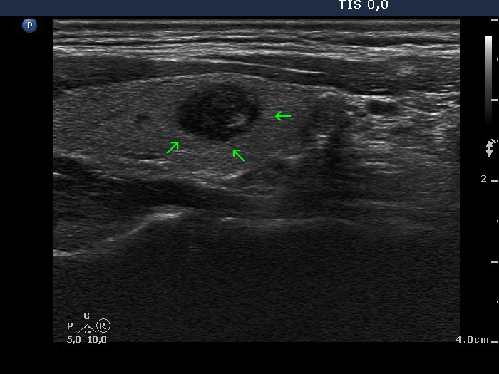
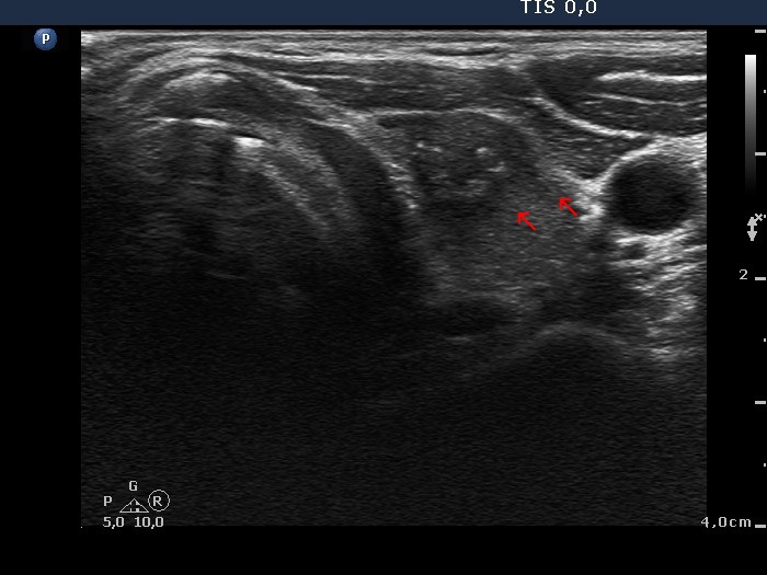
Papillary carcinoma (middle) and benign hyperplastic nodule (lower) (histology) - case conp034

Transverse scan
Longitudinal scan

The middle, malignant nodule has partly blurred (yellow arrows), partly lobulated margins (white arrows) while the lower, benign nodule presents blurred borders (yellow arrows) and spiculated margins (red arrows), as well. The small irregularities marked with green arrow cannot be judged as pathognomonic for lobulated margins.


Papillary carcinoma (histology) - case conp032

Transverse scan
Longitudinal scan

The dorsal border of the nodule is all along blurred and beside equivocal forms (red arrows) has one protrusion (yellow arrow) diagnostic of lobulated margins.

Benign nodular goiter (cytology) - case 2117
Papillary carcinoma (cytology) - case 2057

There are small irregularities on the nodule' border. In my opinion, the extent of protrusions does not reach the pathological degree.

This is an unequivocal example of lobulated margins.

   
Benign nodular goiter (cytology) - case 2054
Benign nodular goiter (cytology) - case 2056

The nodular area presented in both cases is composed of several (left case) and two (right case) discrete lesions causing a lobular appearance. In fact, the lobules are different lesions in the left case which means that this type of lobulation should not be counte as pathological. On the other hand, the right case presents at least one protrusion (yellow arrow), which might be pathological. (It is worth to analyze the protrusion in the video: compared with the still image, it is much less evident that this protrusion would be pathological. As a rule, video is always superior to still image.)

   
Benign nodular goiter (cytology) - case 2024
Benign nodular goiter (cytology) - case 2140

The nodular area is composed of multiple lesions in both cases which causes a lobular appearance. On the other hand, there are several suspicious protrusions on the margin in the right case, therefore the possibility of lobulated/spiculated margins can be raised in this nodule.

   
Benign nodular goiter (cytology) - case 2107
Benign nodular goiter (cytology) - case 2109

In both cases the nodular area is composed of multiple lesions which makes the appearance of the entire mass lobulated. However, this does not mean that the borders of a nodule are in fact lobular. On the other hand, the margin of the nodular area of a single lesion is spiculated or lobulated (yellow arrows).

Hashimoto's thyroiditis (cytology) - case 1496
Benign nodular goiter (cytology) - case 2050

The Hashimoto's case has numerous discrete lesions presenting lobulated and spiculated margins. These lesions are not pathological nodules but more active foci of the thyroiditis.

There is a nodule in the ventral part of the lobe. The lesion presents both lobulations (yellow arrow) and at least one spiculation (red arrow). The latter is characterized by sharp angles.

Hashimoto's thyroiditis (cytology) - case 1768
Hashimoto's thyroiditis (cytology) - case 2168

Both cases are typical presentations of Hashimoto's thyroiditis: there are numerous discrete hypoechogenic lesions with irregular, partly lobulated, partly spiculated margins within an echonormal background. There is no doubt that the margins are irregular, the issue is the differentiation of hypoechogenic lesions of thyroiditis from pathological nodules. None of these lesions corresponds to pathological nodules.

Hashimoto's thyroiditis (cytology) - case 2168

The two protrusions (marked with red arrows) make the appearance of the margin lobulated. However, these lobulations are caused by the infiltration of the underlying thyroiditis (yellow arrows).

Hashimoto's thyroiditis (cytology) - case 430
Benign nodular goiter (cytology) - case 2109

Multiple hypoechogenic areas are presented. These lesions have irregular, lobulated margins.

There is a nodular area with irregular, partly spiculated margins. The nodular area is located in only one part of the lobe. This arrangement would be unusual in Hashimoto's thyroiditis.

It is worth comparing the extralesional part of the thyroid: in Hashimoto's case this is minimally hypoechogenic and presents small, more hypoechogenic areas while in the right case the extranodular part of the lobe is intact.

Hashimoto's thyroiditis (cytology) - case 880
Benign colloid goiter (cytology) - case 2120

This case presents numerous discrete lesions with identical echogenicity, all lesions are hypoechogenic. The borders are partly irregular (lobulated and spiculated).

A multinodular goiter is demonstrated. In this multinodular goiter the nodules differ in echogenicity. The larger heterogeneous (partly hypoechoic, partly isoechoic) nodule located in the dorsal part has lobulated margins.

There are two important differences between the two cases. First, although the borders of the lesions in the thyroiditis' case do not fulfill the criteria of ill-defined borders, they are less well-demarcated compared with the nodule in the right case. In contrast with the Hashimoto's case in which the lesions are uniformly hypoechoic, the nodules in the multinodular goiter' case present different echo patterns.

Hashimoto's thyroiditis (cytology) - case 479
Benign hyperplastic nodules and Hashimoto's thyroiditis (histology) - case 54

In both cases there is an echonormal area composed of multiple lesions within a hypoechoic background. Although the echonormal lesions seemingly present lobulated margins, the cause of these protrusions might differ from the usual lobulation of a nodule. In Hashimoto's thyroiditis it is a reasonable assumption that not the isoechoic lesion, but the underlying thyroiditis is the infiltrative process, and in fact not the echonormal lesion presents lobulations but the underlying thyroiditis infiltrates the isoechoic part making the surface of the latter lobulated. In other words, the isoechoic projections (red arrows) are secondary to the 'obulations' or 'spiculations' of the thyroiditis (yellow arrows).

Hashimoto's thyroiditis (cytology) - case 2080
Papillary carcinoma (cytology) - case 2057

It is equivocal whether the lobular appearance is caused by the presence of multiple lesions next to each other or this is one large single lesion displaying lobulated margins.

This is an unequivocal example of lobulated margins.

There are two main differences between the two cases. The normal parenchyma is hypoechoic in the left while echonormal in the right case. While the benign lesion might be composed of several discrete lesions, the malignant focus is clearly one lesion.

Subacute, de Quervain's thyroiditis (cytology) - case 1454

The hypoechogenic lesions have spiculated margins.

Papillary carcinoma and Hashimoto's thyroiditis (histology) - case 2138

Papillary carcinoma and Hashimoto's thyroiditis in the right lobe
Hashimoto's thyroiditis in the left lobe

The malignant focus has irregular, lobulated margins as has the hypoechoic lesion in the right image.