Table 8. The issue of thyroiditis

doi: 10.24390/thyrosite.ctu.2.5.lect.08.2

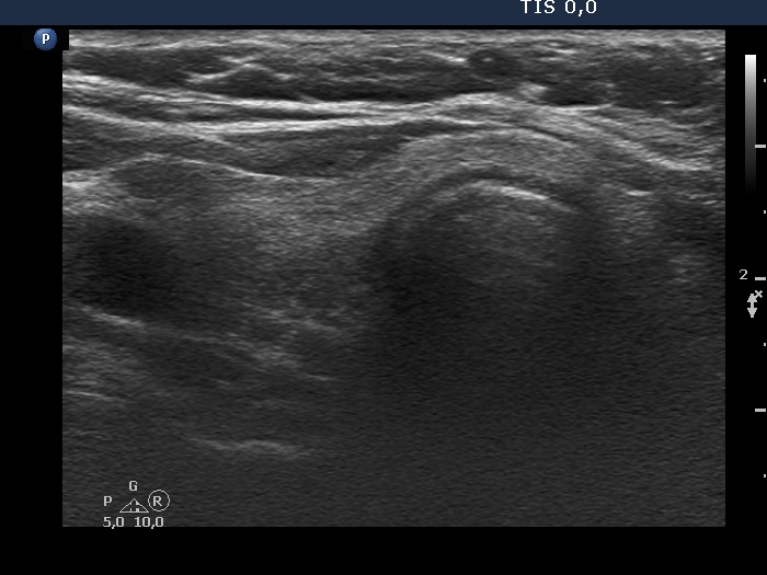
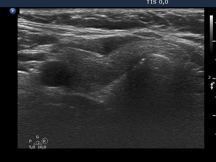
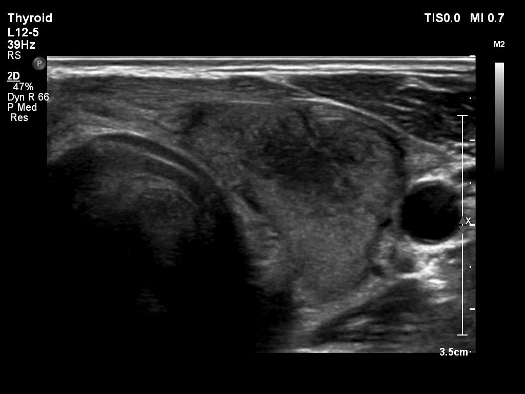
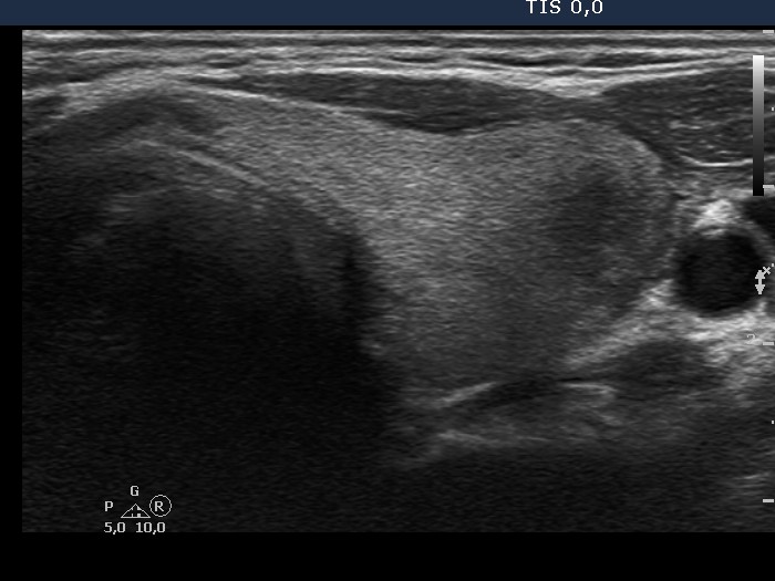
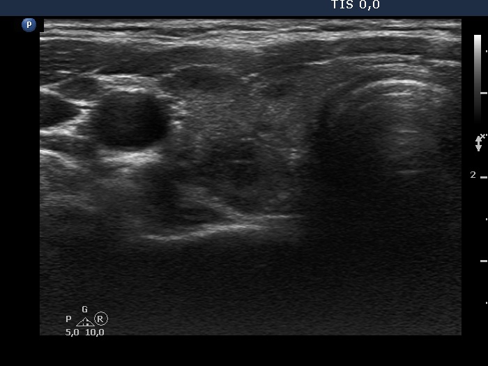
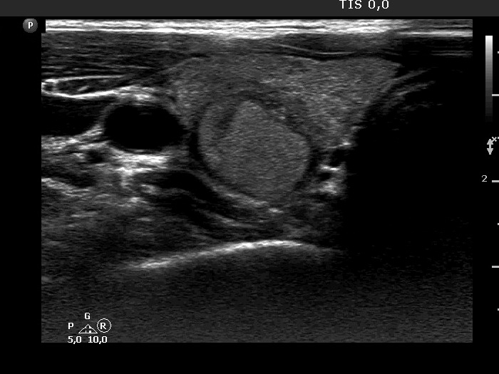
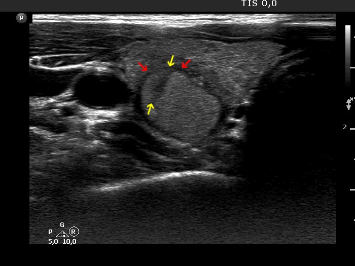
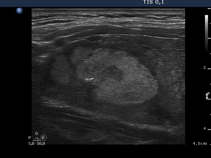
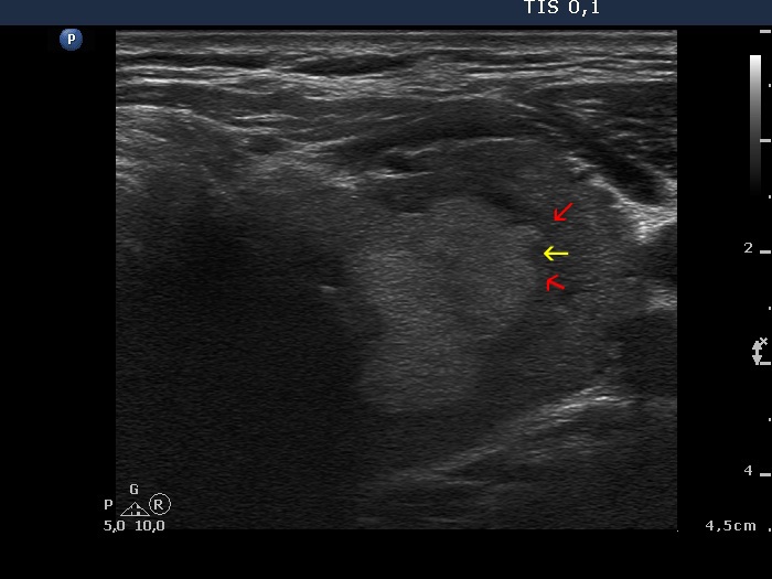
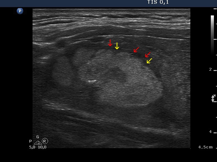
Both Hashimoto's and de Quervain's thyroiditis cause differential diagnostic issue in the judgement of nodule borders. The former is characterized by discrete hypoechoic lesions in more than 90% of cases. These discrete areas usually have blurred or puzzle-like, spiculated margins. The issue is not the presence of abnormal borders but the presence of pathological nodule. We face with another problem in the coexistence of Hashimoto's thyroiditis and pathological nodules. The underlying thyroiditis can infiltrate the nodule and makes the appearance of the nodule lobulated. Therefore, in most of these cases the primary event is the infiltrative process of the thyroiditis, and not that of the nodule. It means that these irregularities of nodule borders should not be encountered among irregular borders.
Both types of thyroiditis frequently present blur of the discrete areas.

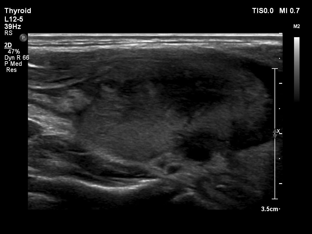
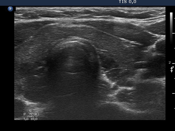
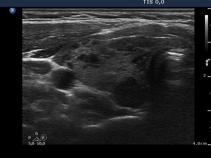
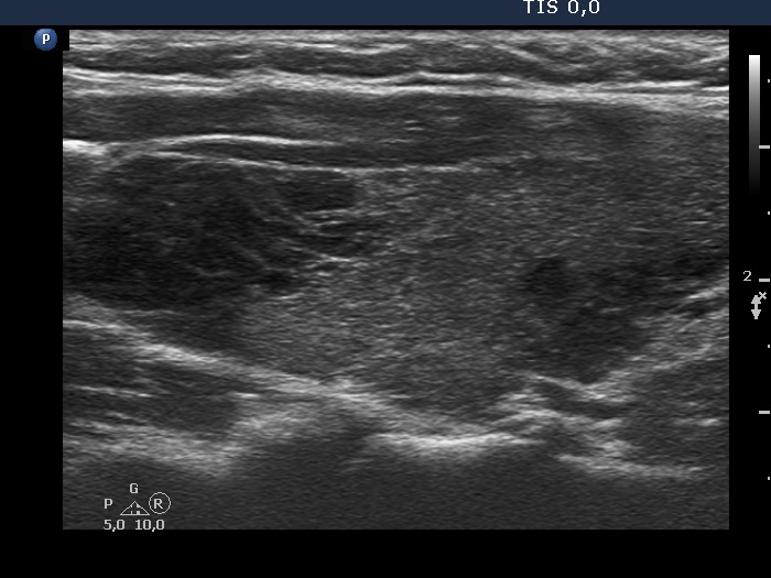
Hashimoto's thyroiditis - case 2108

This case should not cause great problem for an experienced sonographer: this is one of the most common patterns of Hashimoto's thyroiditis. The echogenicity index (the % of hypoechogenic areas) is more than 50%. These hypoechogenic areas have ill-defined borders.

   
Subacute, de Quervain's thyroiditis (cytology) - case 1036
Subacute, de Quervain's thyroiditis (cytology) - case 1454

The borders of the discrete hypoechoic areas are blurred in both cases, in the right case even lobulated. Taking the clinical picture into account, we can avoid overrating this pattern as suspicious for malignancy; in fact, this is the typical presentation of subacute, de Quervain's thyroiditis.

   
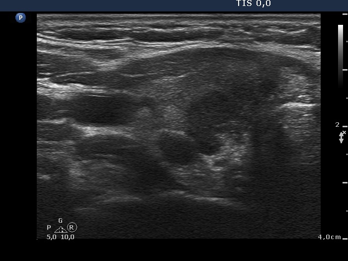
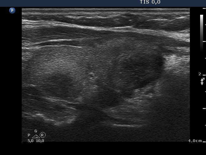
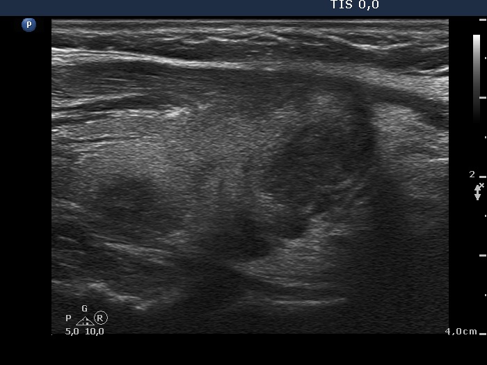
Hashimoto's thyroiditis (cytology) - case 1188
Transverse scans
Longitudinal scans

The thyroid presents numerous hypoechogenic lesions with partly blurred and partly spiculated margins. The presence of multiple hypoechoic lesions argues for Hashimoto's thyroiditis and stands against the presence of pathological nodule.

   
Hashimoto's thyroiditis (cytology) - case 430
Transverse scan
Longitudinal scan

The situation is similar to the former: the thyroid has numerous hypoechoic areas which present irregular borders. The clue is again the recognition that these discrete areas are not pathological nodules but more active foci of Hashimoto's thyroiditis.

   
Hashimoto's thyroiditis (cytology) - case 2080
Hashimoto's thyroiditis (cytology) - case 1496

Both cases present numerous hypoechoic areas with puzzle-like, spiculated margins. In fact, this is one of the most typical presentations of Hashimoto's thyroiditis.

   
Hashimoto's thyroiditis (cytology) - case 2168

The two protrusions (marked with red arrows) make the appearance of the margin lobulated. However, these lobulations are caused by the infiltration of the lesion by the underlying thyroiditis (yellow arrows).

   
Hashimoto's thyroiditis (cytology) - case 54

The primary protrusions in this case are caused by the underlying thyroiditis (yellow arrows), while the protrusions of the isoechoic lesion marked with red are only consequences of the former.