|
|
 |
 |
 |
 |
This comparison points out why the importance of bulging should be treated with caution. The left case presents bulging of the thyroid' contour caused by a nodule which contour is not bulging because it is covered by normal tissue. On the other hand, the nodule in the right case presents bulging contours. The difference might be caused either by the invasive nature of a carcinoma or simply by the location of the nodule.
|
| |
|
|
 |
 |
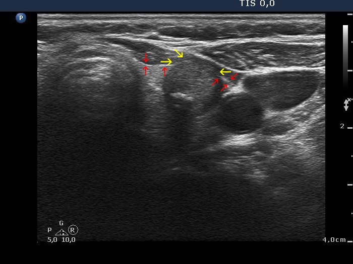
Red arrows point to the most ventral part of the lobe outside the nodule while yellow arrows do to the most ventral part of the tumor. The difference means an unusually large degree of bulging.
|
| |
|
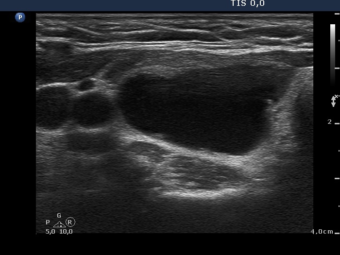
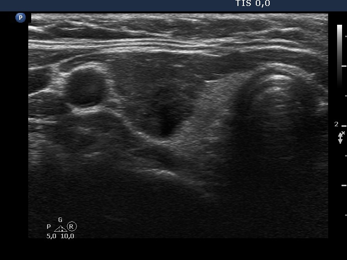
Sclerotherapy of a benign cystic nodule - case 2068 |
Before the second session of sclerotherapy |
Four years after the sclerotherapy |
 |
 |
 |
 |
Initially, the nodule presented non-abutting contour because there was a thin echonormal parenchyma between the ventral surface of the nodule and the thyroid. At the follow-up, the contour became abutting. The capsule was non-visible at the first, while became discontinuous at the follow-up examination.
|
| |
|
|
 |
 |
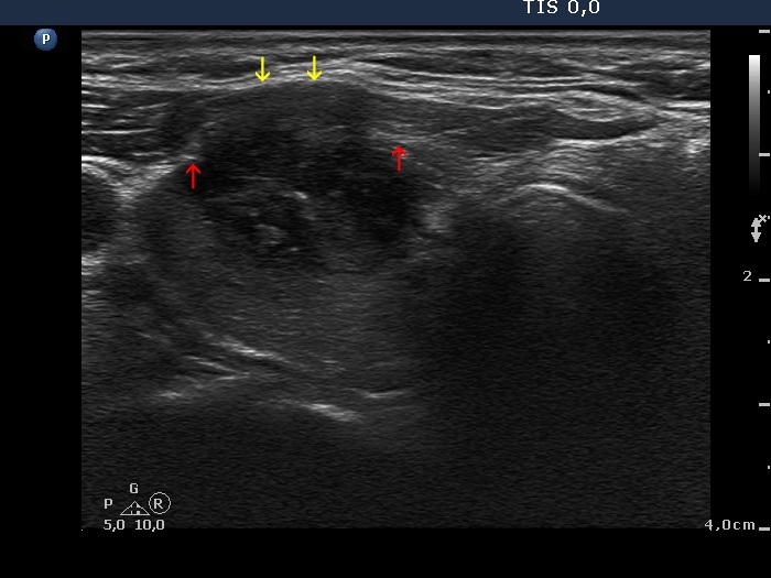
The contour of the lobe is marked with yellow arrows, the contour of the tumor is marked with red arrows. Due to the bulging of the tumor, the sternocleidomastoid muscle became narrow. The normal part of the muscle is marked with white arrows.
|
| |
|
|
Transverse scan |
Longitudinal scan |
 |
 |
 |
 |
The hyperechogenic line on the ventral surface of the lobe is all along intact in the transverse scan, while the more complex echogenic structure seen in the longitudinal scan is not broken. Note that the contour is bulging on the transverse scan.
|
| |
|
|
Transverse scan |
Longitudinal scan |
 |
 |
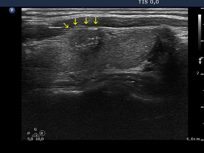
The nodule has macrocalcifications and therefore shows acoustic shadow. The latter hinders the judgment of all signs on extrathyroidal spread.
|
| |
|
|
 |
|
 |
 |
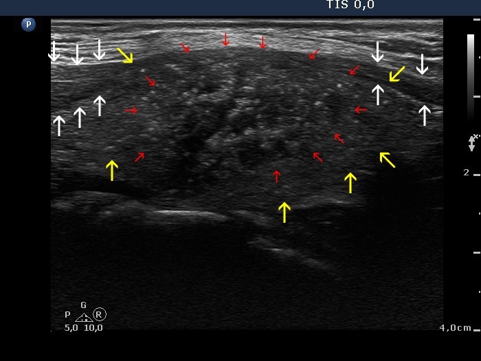
There is an area where we cannot separate the strap muscle from the cystic nodule. This is caused simply by a technical artifact: be aware that we can follow an acoustic artifact (marked with red) up to the most ventral part of the image.
On the other hand, this case meets all criteria on which extrathyroidal spread should be considered: abutment, protrusion of the nodule into the adjacent tissue (i.e. capsular bulging) and disruption of the capsule (green arrows).
|
| |
|
|
Transverse scan |
Longitudinal scan |
 |
 |
 |
 |
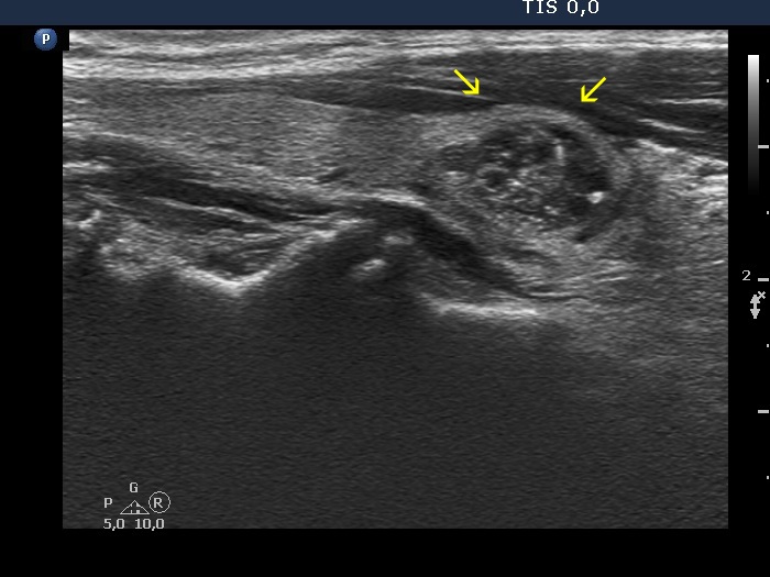
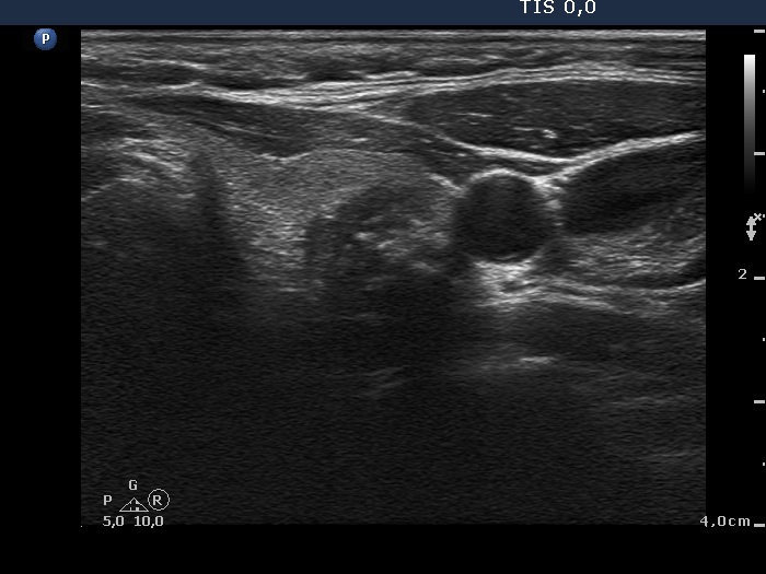
The nodule presents all three signs on which extrathyroidal extension has to be considered: the contour is abutting and bulging while the capsule is discontinuous at the abutting portion of the nodule (yellow arrows).
|
| |
|
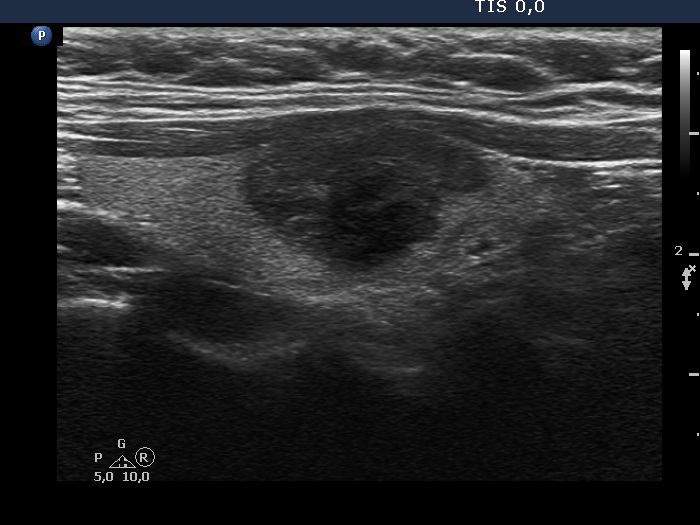
Benign case, an autonomously functioning adenoma - case 2069 |
 |
 |
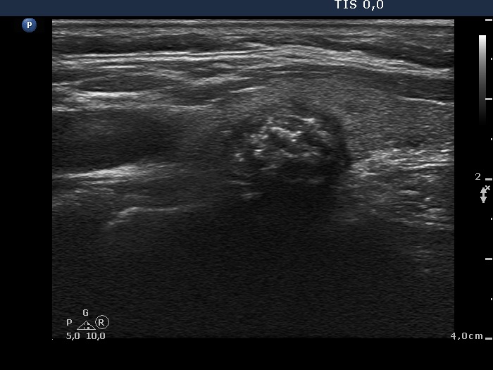
The contour is bulging in this case, however, the nodule is located in the ventromedial, isthmic part of the lobe: nodules is such localization are almost always bulging simply because of the anatomical situation.
|