Intranodular hyperechogenic figures - Table 2 (large). Comet-tail artifacts or colloid crystals |
||
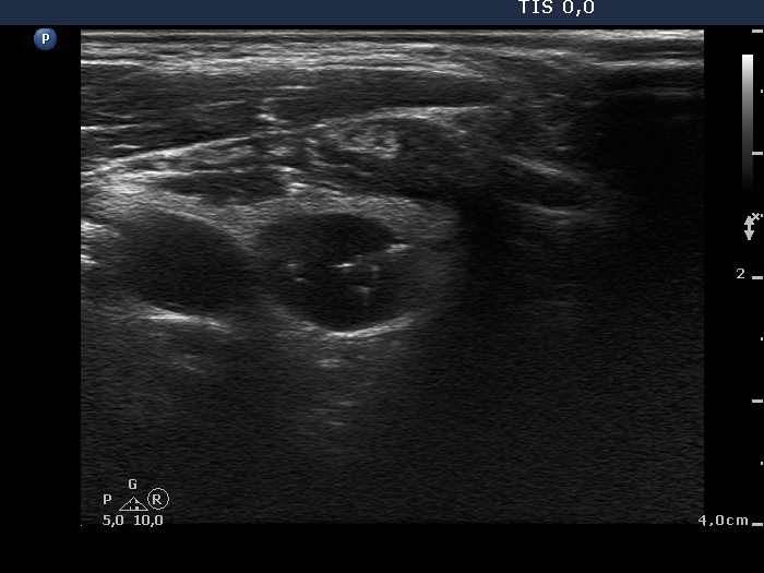
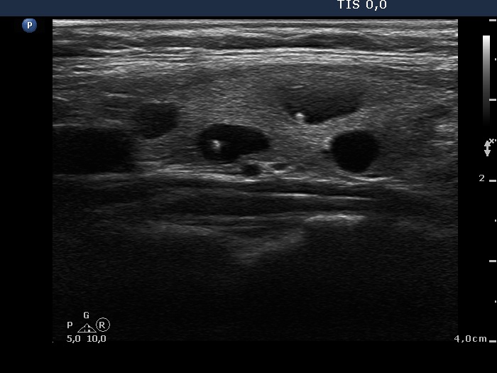
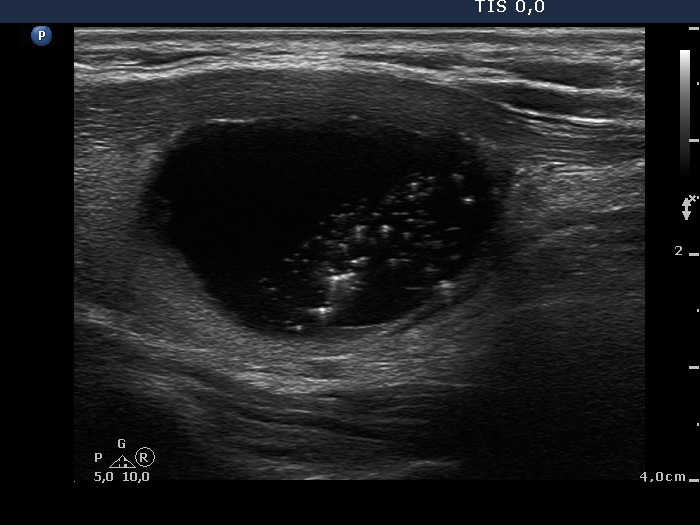
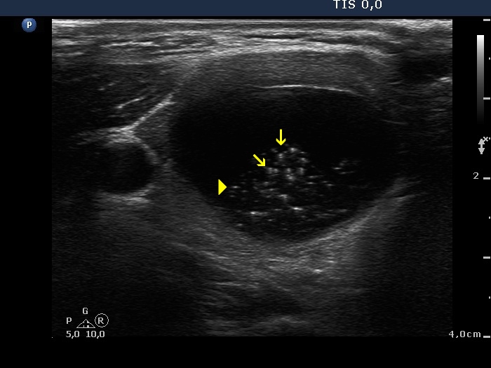
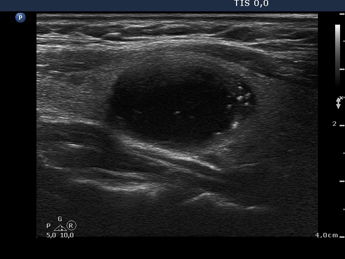
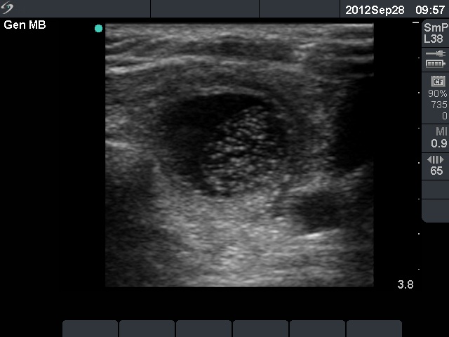
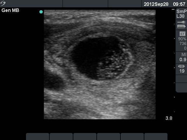
A typical comet-tail artifact consists of a hyperechogenic granule with a dorsal narrowing and a fading tail. The size of the granule is in the range of punctate echogenic foci (microcalcifications), i.e. not greater than 1 mm. The typical presentation of this figure is best seen in cystic part of a mixed nodule; however it might occur within the solid area, as well. In the latter case the narrowing tail is frequently missing.
Benign cystic-colloid goiter (cytological diagnosis) - case 284 |
|
 |
 |
|
 |
|
|
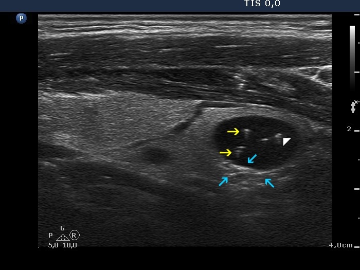
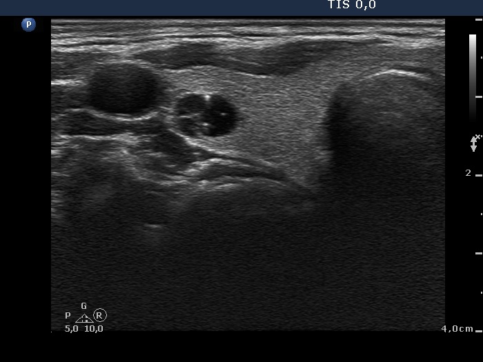
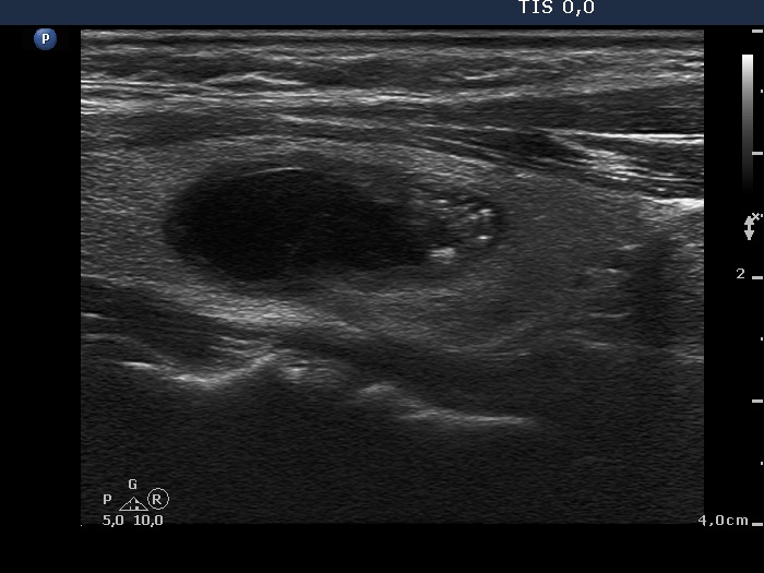
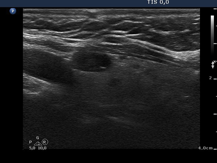
Benign follicular proliferation (cytological diagnosis) - case 270 |
|
 |
 |
A typical comet-tail artifact is demonstrated. |
|
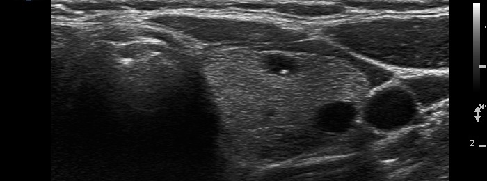
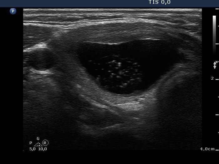
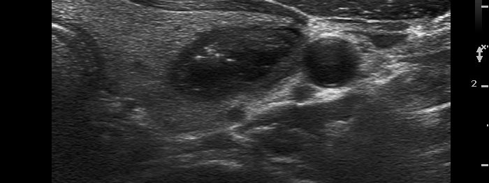
Intact thyroid with cystically dilated macrofolliculi (histological diagnosis) - case 1539 |
|
 |
 |
|
|
  |
 |
Left lobe: there are typical comet-tail artifacts in this lobe, too. |
|
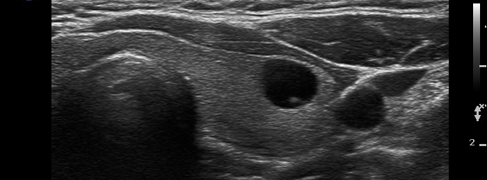
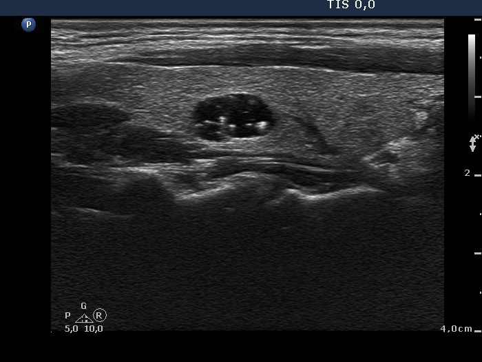
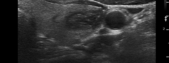
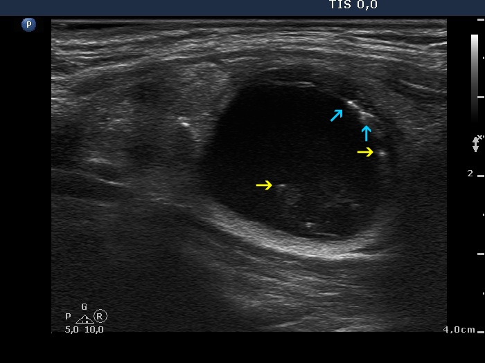
Benign colloid cyst (cytological diagnosis) - case 402 |
|
 |
 |
 |
 |
There are several typical comet-tail artifacts in both lobes.
|
|
Benign cystic degeneration (cytological diagnosis) - case 624 |
|
 |
 |
 |
 |
|
 |
|
|
Follicular adenoma (histological diagnosis) - case 443 |
|
Before aspiration of 2 mL cystic fluid |
|
Upper and lower transverse section |
Longitudinal section |
  |
 |
|
|
After aspiration of 2 mL cystic fluid |
|
Upper and lower transverse section |
Longitudinal section |
  |
 |
|
|
Hashimoto's thyroiditis with several cystic areas but without any nodules (histological diagnosis) - case 1365 |
|
 |
 |
|
|
Benign colloid goiter (cytological diagnosis) - case 386 |
|
 |
 |
|
|
Benign cystic degeneration (cytological diagnosis) - case 662 |
|
 |
 |
|
|