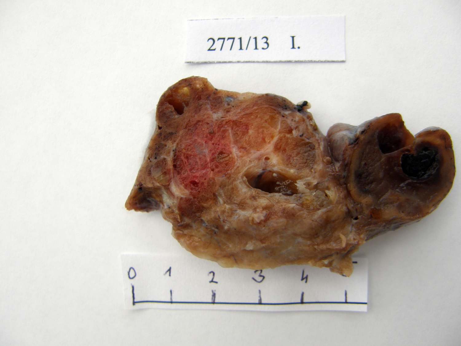
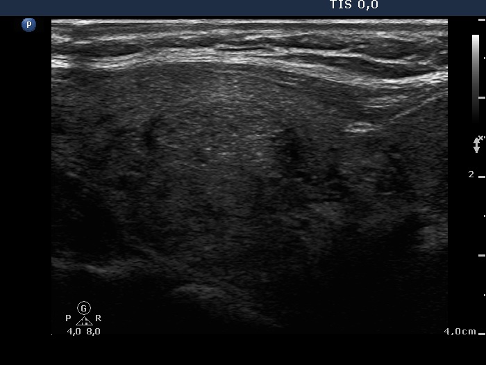
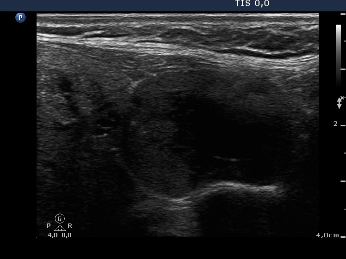
Macroscopic presentation in comparison with ultrasound images - Figure 10. Coexistent benign hyperplastic nodules and a follicular adenoma.
| The upper two third of the lobe (left two-third in the image) on macroscopic photo is presented in ultrasound image. This part of the thyroid is composed of numerous hyperplastic nodules. | |
 |
 |
The lower third of the lobe (right third in the image) on macroscopic photo is presented in ultrasound image. This part of the thyroid contains a nodule presenting capsule. Thie lesion proved to be a follicular adenoma. |
|
|
 |