
Follicular adenoma - Case 8. (ultrasonographic picture 6)
|
|

|
|
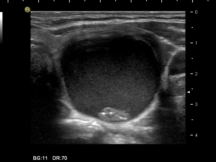
Left lobe, horizontal scan. There is a large cystic nodule. The solid part of the lesion contains bright echogenic granules. These are related to ventral cystic areas, therefore these granules are probably back wall figures. Nevertheless, the possiblity of microcalcifications cannot be fully excluded.