
|
|
Follicular adenoma - Case 28.
|
|


Clinical data: a 40-year-old man with a nodule discovered 6 months earlier.
Palpation: the left lobe was enlarged, and a moderately firm nodule was palpable within the lobe.
Functional state: euthyroidism with TSH-level 1.90 mIU/L.
Ultrasonography: a moderately hypoechogenic nodule comprising greater part of the left lobe. A halo sign is not present on gray scale mode. The presence of perinodular blood flow is equivocal.
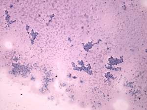
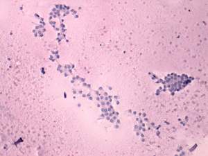
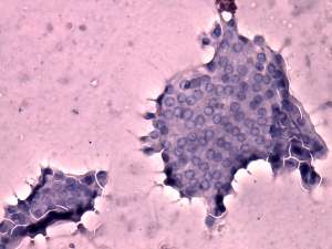
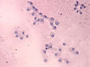
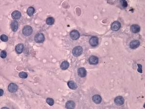
Cytological picture: diffuse colloid precipitate. Thyrocytes are arranged predominantly in normo- and microfollicles. These are regular. Several thyrocytes exhibit nuclear groove and inclusion. In a few clusters, the nuclei of the follicular cells are pale.
Cytological diagnosis: follicular tumor with great probability, but papillary cancer cannot be excluded.
Histopathology: revealed normofollicular adenoma.