
|
|
Follicular adenoma - Case 9.
|
|


First examination (first and second row of images):
Clinical data: a 29-year-old woman was referred for an evaluation of a "cold" nodule. She was examined because of infertility.
Palpation: no abnormality.
Functional state: euthyroidism with TSH 0.55 mIU/L.
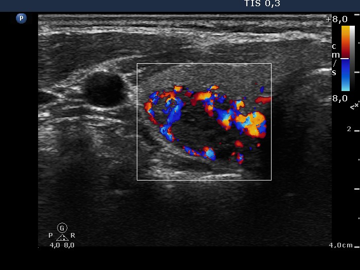
Ultrasonography: a hypoechogenic nodule displaying a type 3 vascular pattern without a halo sign. The dimensions of the nodule were 13x9x15 mm.
Cytology resulted in suspicion of a follicular tumor.
Considering the lack of halo sign and perinodular blood flow and the lack of cytologial atypia we offered follow-up examinations instead of an immediate surgery.
Second examination 2 years later (third and fourth row of images):
Clinical data, palpation and functional state: unchanged. The TSH was 0.72 mIU/L.
Ultrasonography: the pattern was unchanged. The dimensions of the nodue were 15x10x18 mm.
Cytology resulted in suspicion of a follicular tumor.
Considering the significant increase in size we advised surgery. The patient refused our suggestion.
Third examination another 7 years later (fifth and sixth row of images):
Clinical data, palpation and functional state: unchanged. The TSH was 0.49 mIU/L.
Ultrasonography: in contrast with previous examinations perinodular blood flow could be detected. The dimensions of the nodule were 16x13x24 mm.
Cytology resulted in suspicion of a follicular tumor
Histopathology disclosed follicular adenoma.