|
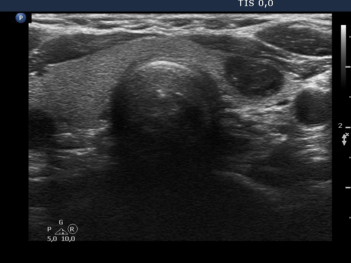
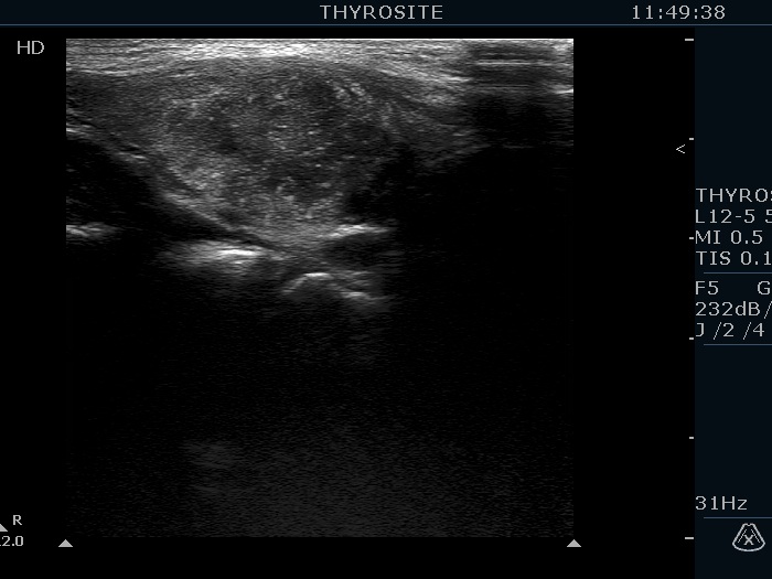
Transverse scan |
 |
 |
Longitudinal scan
|
 |
 |
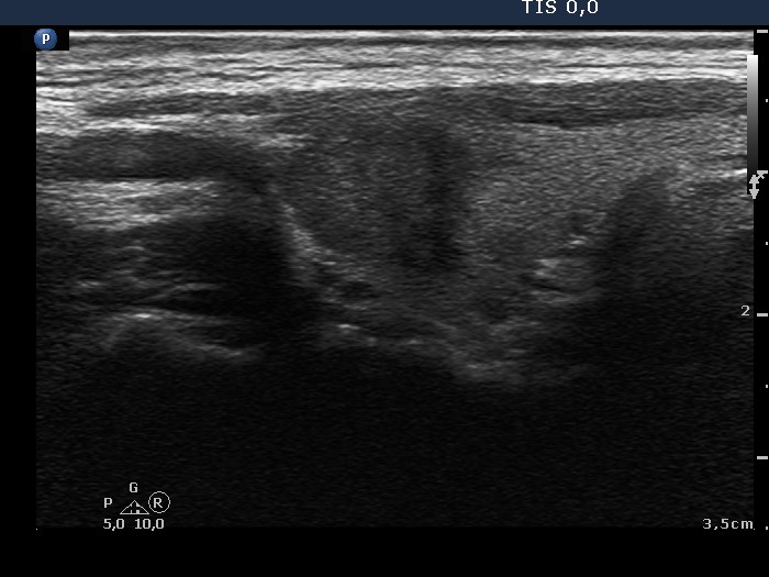
The ventral part of the nodule is ill-defined on longitudinal scan. However, even in these images the degree of blur is below 50%. Moreover, the undefined margins are mainly caused by the identical echogenicity of the ventral part of the lobe (yellow arrow) and the neighboring strap muscle (red arrow). The small irregularities marked with green do not reach the extent of pathological degree.
|
| |
|
|
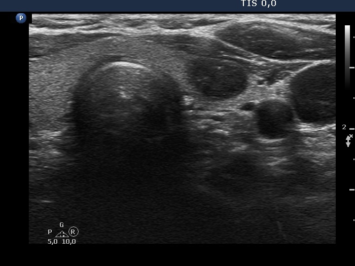
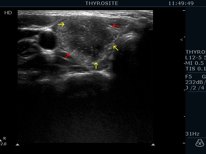
Transverse scan |
Longitudinal scan |
 |
|
 |
 |
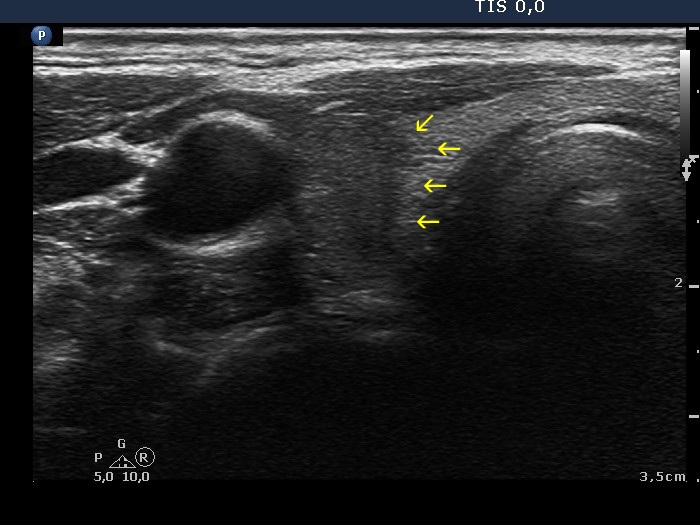
Three types of indistinct borders are presented in these images. Green arrows point to that part of the nodule which echogenicity is identical to the extrathyroidal strap muscle. The tumor has partly blurred (red arrows) and partly spiculated (yellow arrows) margins. The extent of blurred part of the border exceeds 50%.
|
| |
|
|
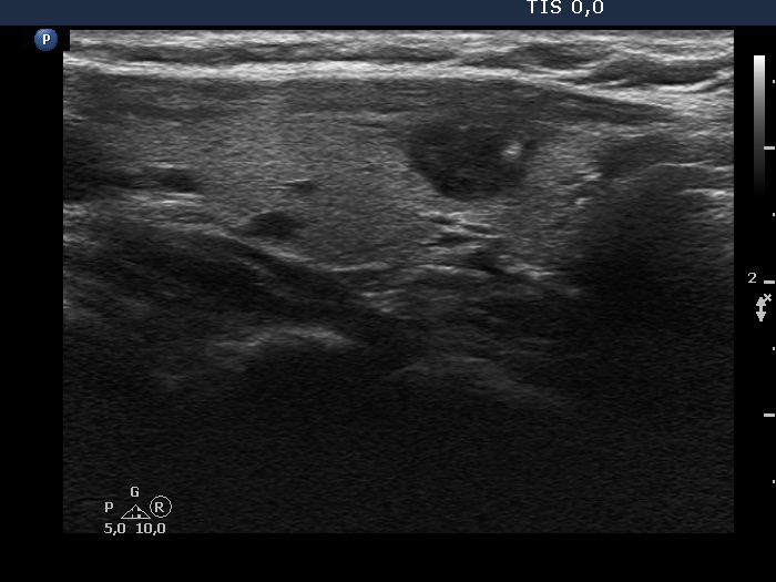
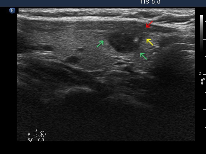
Transverse scan |
Longitudinal scan |
 |
 |
 |
 |
In this case almost the entire border of the nodule is ill-defined, moreover the lesion has lobulated (yellow arrows) and spiculated (red arrows) margins, as well.
|
| |
|
|
Transverse scan |
Longitudinal scan |
 |
 |
In this case more than 50% of the nodule' borders are ill-defined.
|
|
| |
|
|
Transverse scan |
Longitudinal scan |
 |
 |
 |
 |
The nodule has ill-defined borders in less than the third and more than the three-fourth of the circumference of the nodule, transverse scan and longitudinal view, respectively. (See the border between the red arrows.) The nodule also presents lobulation, the small irregularities marked with while arrow are not necessarily pathological, while those marked with yellow are abnormal findings.
|
| |
|
|
Transverse scan |
Longitudinal scan |
 |
 |
The borders of the nodule are both blurred and spiculated-lobulated.
|
|
| |
|
|
Transverse scan |
Longitudinal scan |
 |
 |
This is a typical example of ill-defined borders.
|
| |
|
|
Transverse scans |
Longitudinal scans |
 |
 |
 |
 |
There are two lesions in the right lobe, a ventral deeply hypoechogenic nodule and a dorsal minimally/moderately hypoechogenic area. Both have blurred borders, the ventral one has spiculated margins, as well.
|
| |
|
|
Transverse scan |
Longitudinal scan |
 |
 |
 |
 |
The dorsal and medial borders of the nodule are partly ill-defined, partly lobulated. |
|
| |
|
|
Transverse scan |
Longitudinal scan |
 |
 |
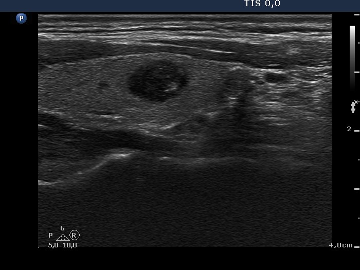
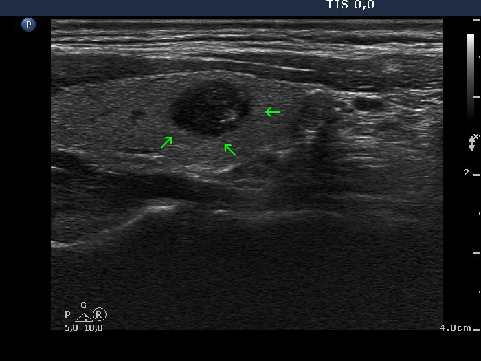
The dorsal part of the nodule is ill-defined.
|
|
| |
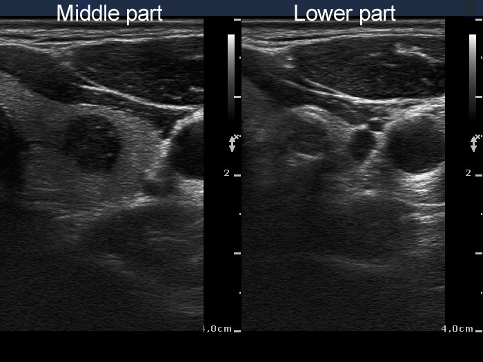
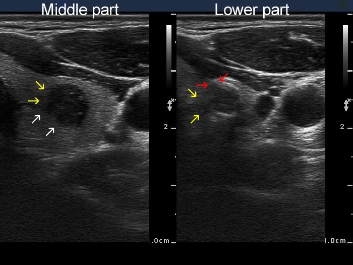
Papillary carcinoma (middle) and benign hyperplastic nodule (lower) (histology) - case conp034
|
Transverse scan |
Longitudinal scan |
 |
 |
 |
 |
The middle, malignant nodule has partly blurred (yellow arrows), partly lobulated margins (white arrows) while the lower, benign nodule presents blurred borders (yellow arrows) and spiculated margins (red arrows), as well. The small irregularities marked with green arrow cannot be judged as pathognomonic for lobulated margins.
|
| |
|
