|
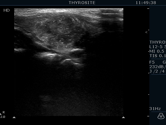
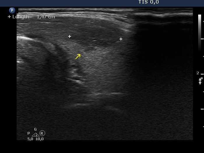
Transverse scan |
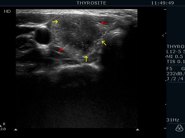
Longitudinal scan |
 |
 |
 |
 |
Three types of indistinct borders are presented in these images. Green arrows point to that part of the nodule which echogenicity is identical to the extrathyroidal strap muscle. The tumor has partly blurred (red arrows) and partly spiculated (yellow arrows) margins.
|
|
|
|
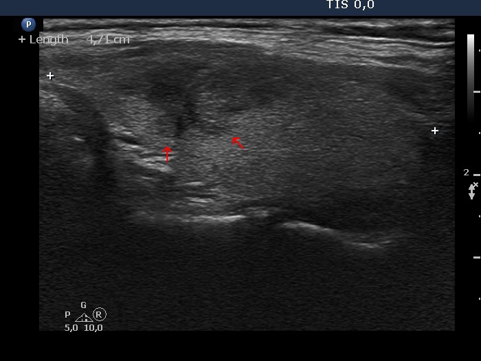
Transverse scan |
Longitudinal scan |
 |
 |
 |
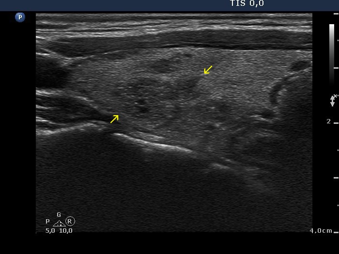 |
The tumor spreads into the normal parenchyma with elongated projections which have sharp edges (yellow arrows).
|
|
|
Papillary carcinoma (upper) and benign hyperplastic nodule (lower) (histology) - case conp027
|
Transverse scan |
Longitudinal scan |
 |
 |
 |
 |
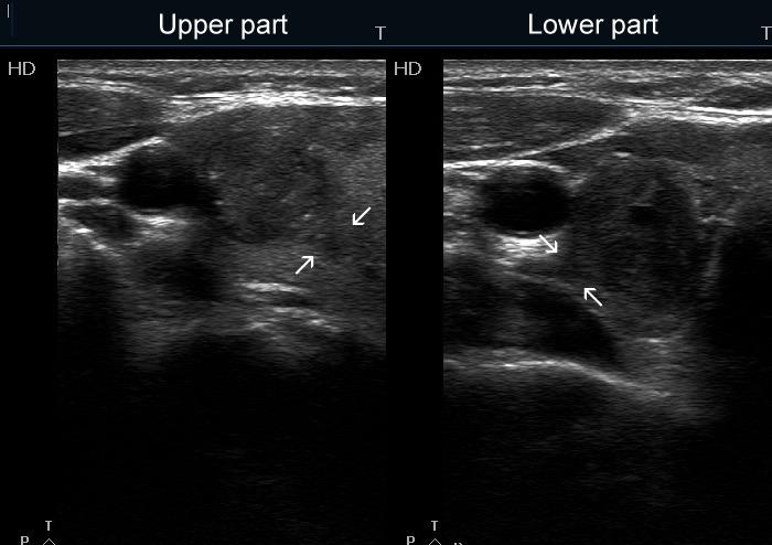
The nodules present different types of pathological borders. White arrows point to spiculated margins, red arrows do to lobulated margins while yellow ones do to blurred borders. The origin of the spiculations on the surface of the hypoechogenic nodule (green arrows) is equivocal, these might be caused by the impression of the echonormal lesion on the surface. The upper, malignant nodule has microcalcifications while the lower benign one presents both taller-than-wide and longer-than wide sign.
|
|
|
Papillary carcinoma (upper) and benign hyperplastic nodule (lower) (histology) - case conp037
|
|
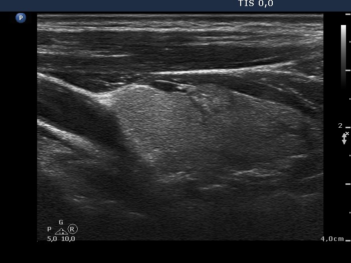
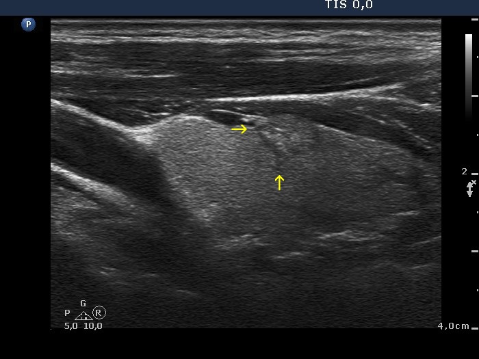
Longitudinal scan
|
 |
 |
The yellow arrows point the spiculated margins.
|
|
|
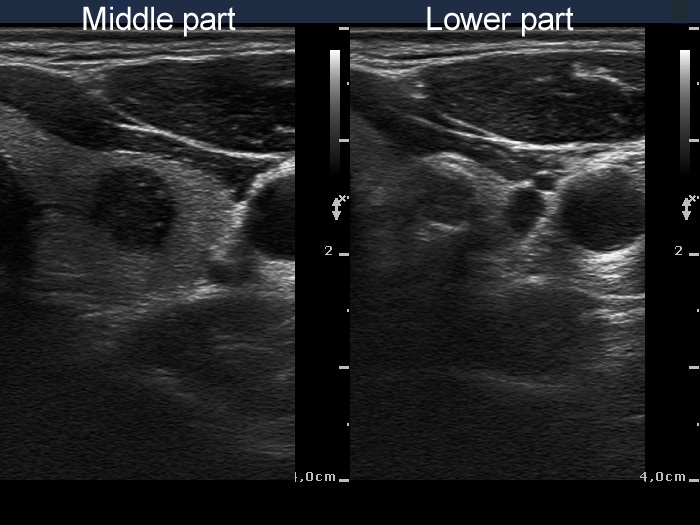
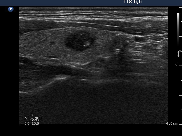
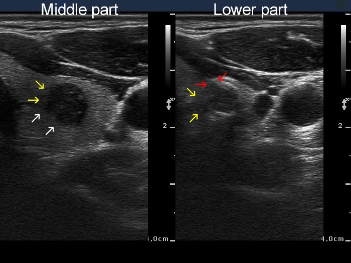
Papillary carcinoma (middle) and benign hyperplastic nodule (lower) (histology) - case conp034
|
Transverse scan |
Longitudinal scan |
 |
 |
 |
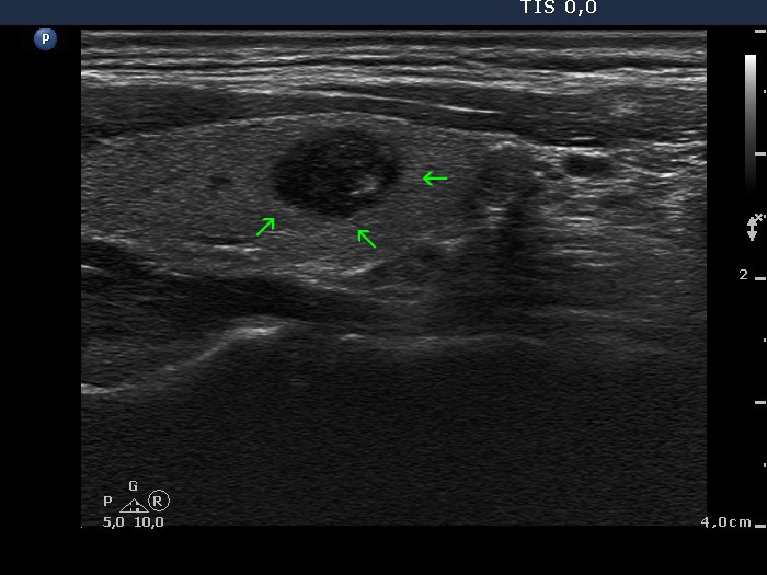 |
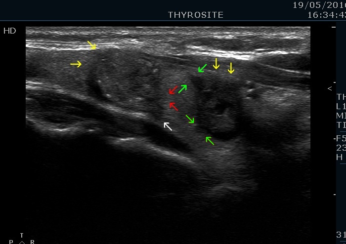
The middle, malignant nodule has partly blurred (yellow arrows), partly lobulated margins (white arrows) while the lower, benign nodule presents blurred borders (yellow arrows) and spiculated margins (red arrows), as well. The small irregularities marked with green arrow cannot be judged as pathognomonic for lobulated margins.
|
|
|
|
Transverse scan |
Longitudinal scan |
 |
 |
 |
 |
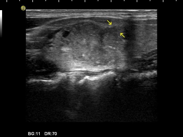
In this case almost the entire border of the nodule is ill-defined, moreover the lesion has lobulated (yellow arrows) and spiculated (red arrows) margins, as well.
|
|
|
|
|
Longitudinal scans |
 |
 |
The nodule has spiculated margins, the dorsal spicule is pathognomonic, the ventral one is less evident.
|
|
|
Oxyphilic variant of papillary carcinoma in an autonomously functioning nodule (histology) - case 2022
|
Transverse scans |
Longitudinal scans |
 |
 |
 |
 |
The nodule has irregular borders, there are some spiculations with sharp angles (yellow arrows) and some protrusions marked with red arrow.
|
| |
|
| |
|
|
|
