
The borders of the nodule - case 1036
30 months after the initial examination (ultrasonographic picture 6)
|
|

|
|
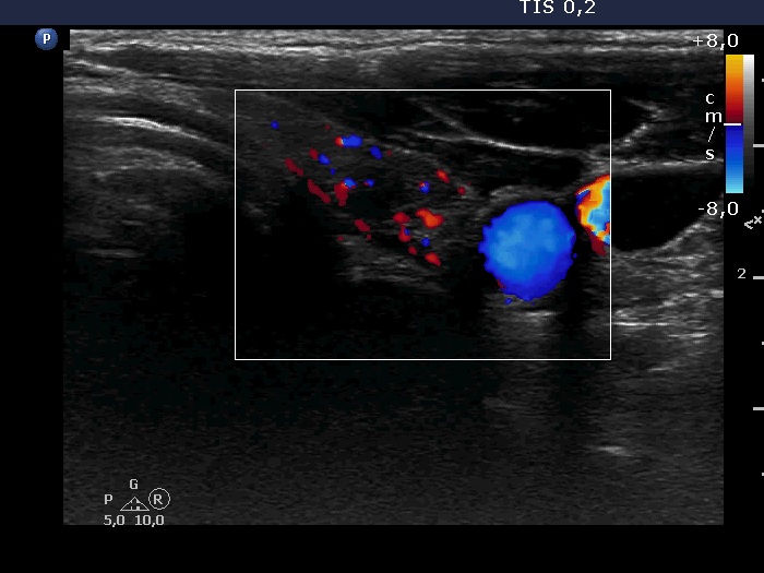
Left lobe, transverse scan, color Doppler mode. The vascularization is a little bit increased.