
The borders of the nodule - case 1454
12 weeks after the initial examination (ultrasonographic picture 5)
|
|

|
|
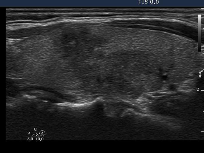
Left lobe, longitudinal scan. This is a typical presentation of de Quervain's thyroiditis: hypoechogenic areas with blurred borders within echonormal background.