The borders of the nodule - case 186 (ultrasonographic picture 2b)
|
|

|
|
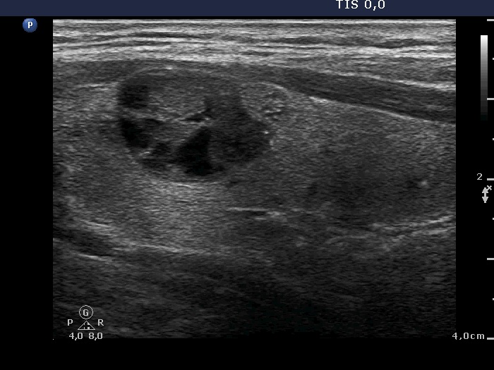
Right lobe, longitudinal view. The lower-ventral part of the hypoechogenic nodule seems to lobulated or even spiculated.

2022-23 Advanced
Papillon Course
Nodule' borders
|
|
|
|
Right lobe, longitudinal view. The lower-ventral part of the hypoechogenic nodule seems to lobulated or even spiculated.