
The borders of the nodule - case 186 (ultrasonographic picture 7)
|
|

|
|
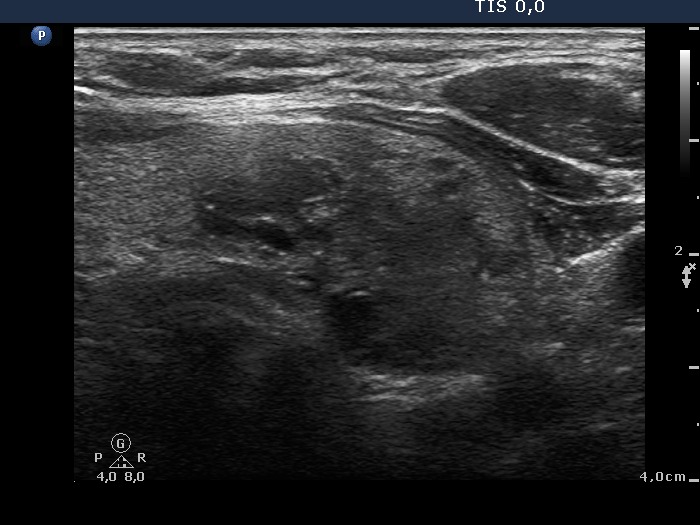
Lower part of the left lobe, transverse scan. There are two moderately hypoechogenic nodules next to each other. They present echogenic figures, both lines and granules. It is more likely that these figures are not microcalcifications but presentations of connective tissue or caused by back wall posterior enhancement.