The borders of the nodule - case 2050 (ultrasonographic picture 7)
|
|

|
|
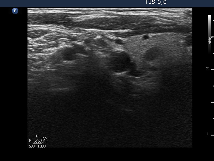
Lateral to the right lobe, longitudinal scan. This hypoechoic mass could be either a lymph node or a muscle fiber.

2022-23 Advanced
Papillon Course
Nodule' borders
|
|
|
|
Lateral to the right lobe, longitudinal scan. This hypoechoic mass could be either a lymph node or a muscle fiber.