
The borders of the nodule - case 2089 (ultrasonographic picture 1)

|
|
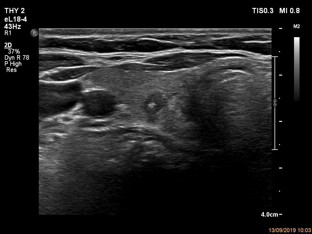
Right lobe, transverse scan. The hypoechoic lesion has microcalcifications, presents taller-than-wide shape. The margins are not regular, but the extent of the irregularity does not reach the degree of pathological level. The dorsal tiny figure may be a distinct, smaller lesion.