
The borders of the nodule - case 2115 (ultrasonographic picture 9)
|
|

|
|
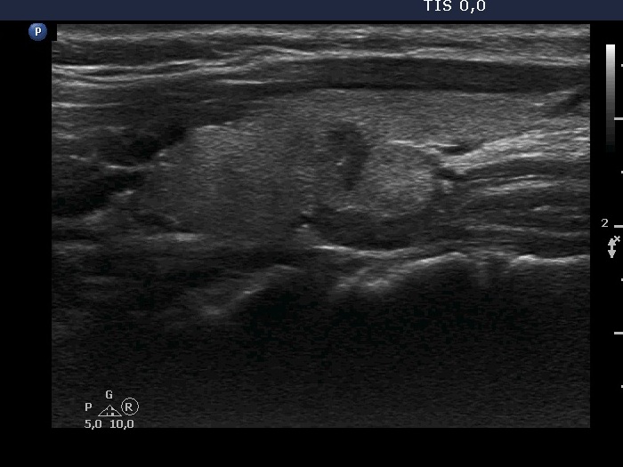
Left lobe, another longitudinal view. On this view, it seems to be more likely that this mass is composed of a hypoechogeninc and an echonormal nodule.