
The borders of the nodule - case 2168 (ultrasonographic picture 6)
|
|

|
|
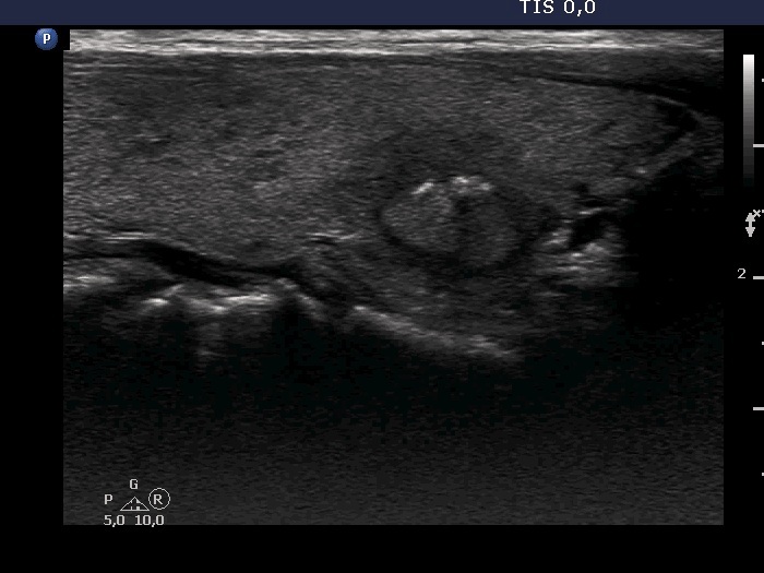
Right lobe, another longitudinal view. These figures are partly microcalcifications and based on the acoustic shadow partly macrocalcification.