
The borders of the nodule - case 2169 (ultrasonographic picture 5)
|
|

|
|
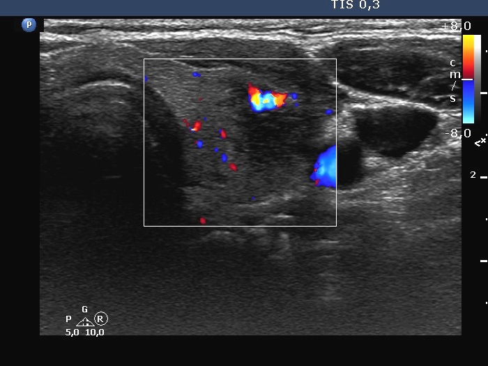
Left lobe, transverse scan, color Doppler mode. Although there is a large vessel within the hypoechoic area, the lesion has a decreased vascularization.