The borders of the nodule - case 349 (picture 2 of intraoperative imprint smear)
|
|

|
 |
|
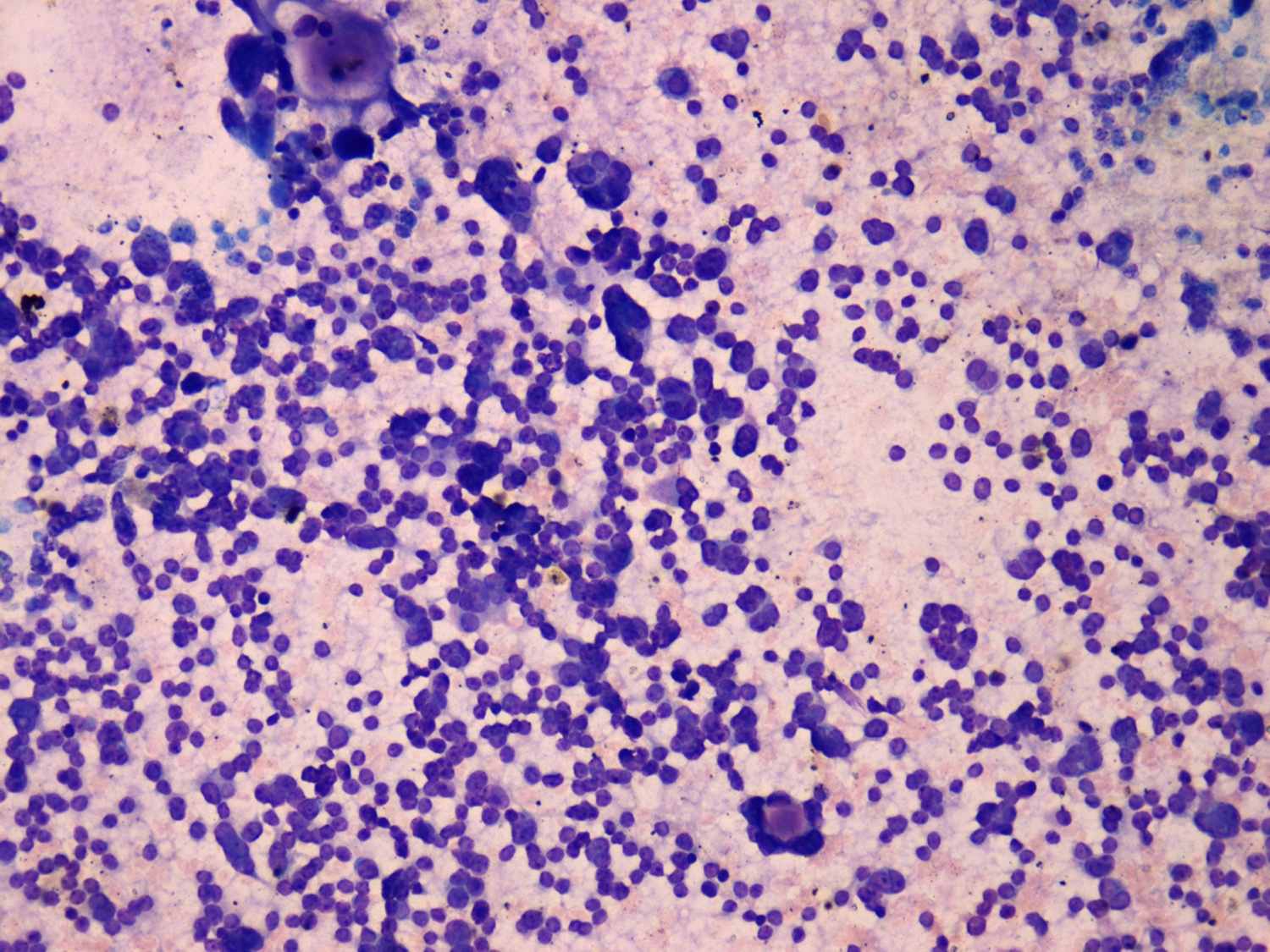
Wright-Giemsa staining, 200x. Dispersed cells predominate the smear. Note a few rosette-like structures.

2022-23 Advanced
Papillon Course
Nodule' borders
|
|
|
|
|
Wright-Giemsa staining, 200x. Dispersed cells predominate the smear. Note a few rosette-like structures.