
The borders of the nodule - case 349 (cytologic picture 6)
|
|

|
 |
|
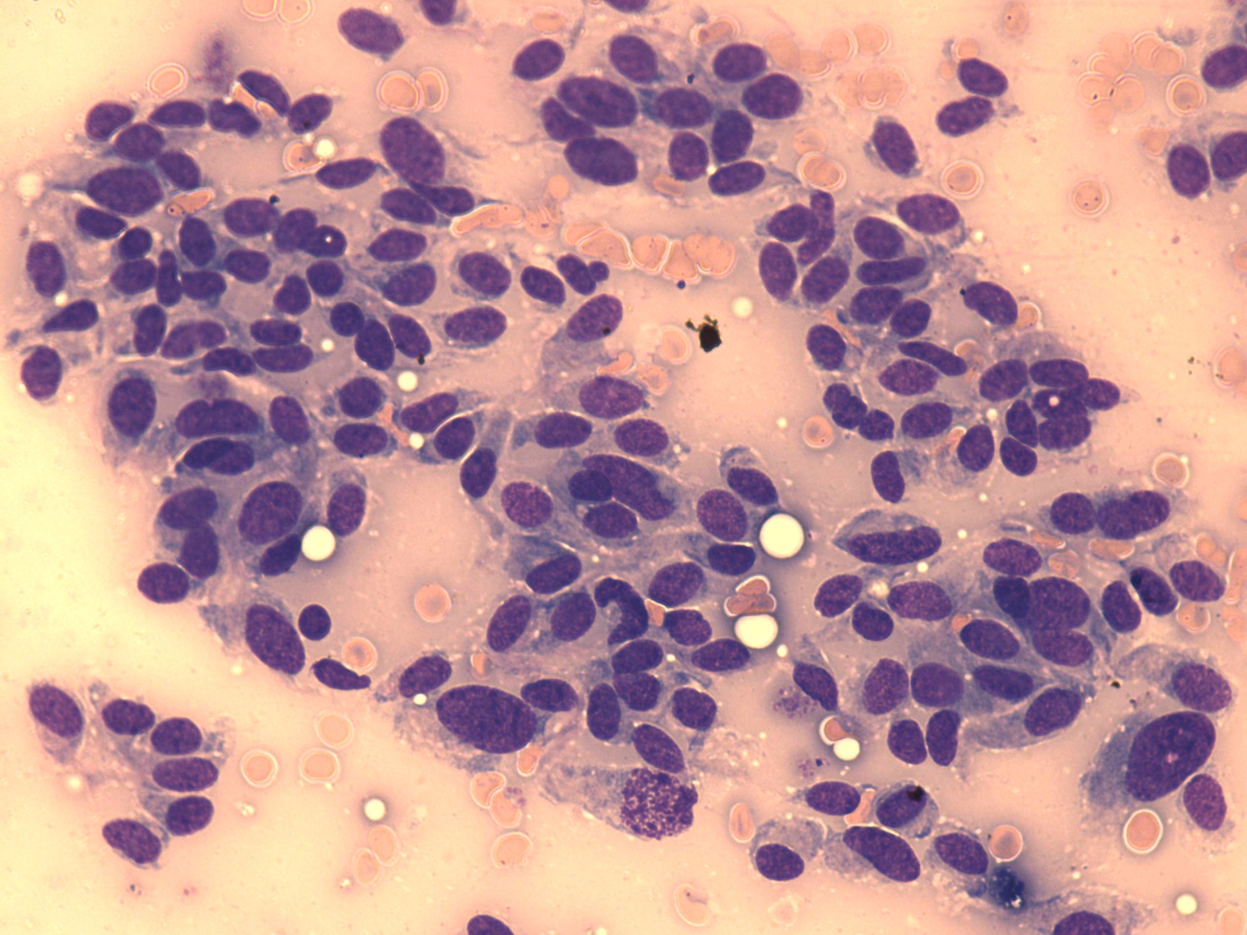
Wright-Giemsa-staining, 400x. Elongated tumor cells predominate the smear. Note a single cell necrosis in the lower part of the image.