
The borders of the nodule - case 349 (cytologic picture 8)
|
|

|
 |
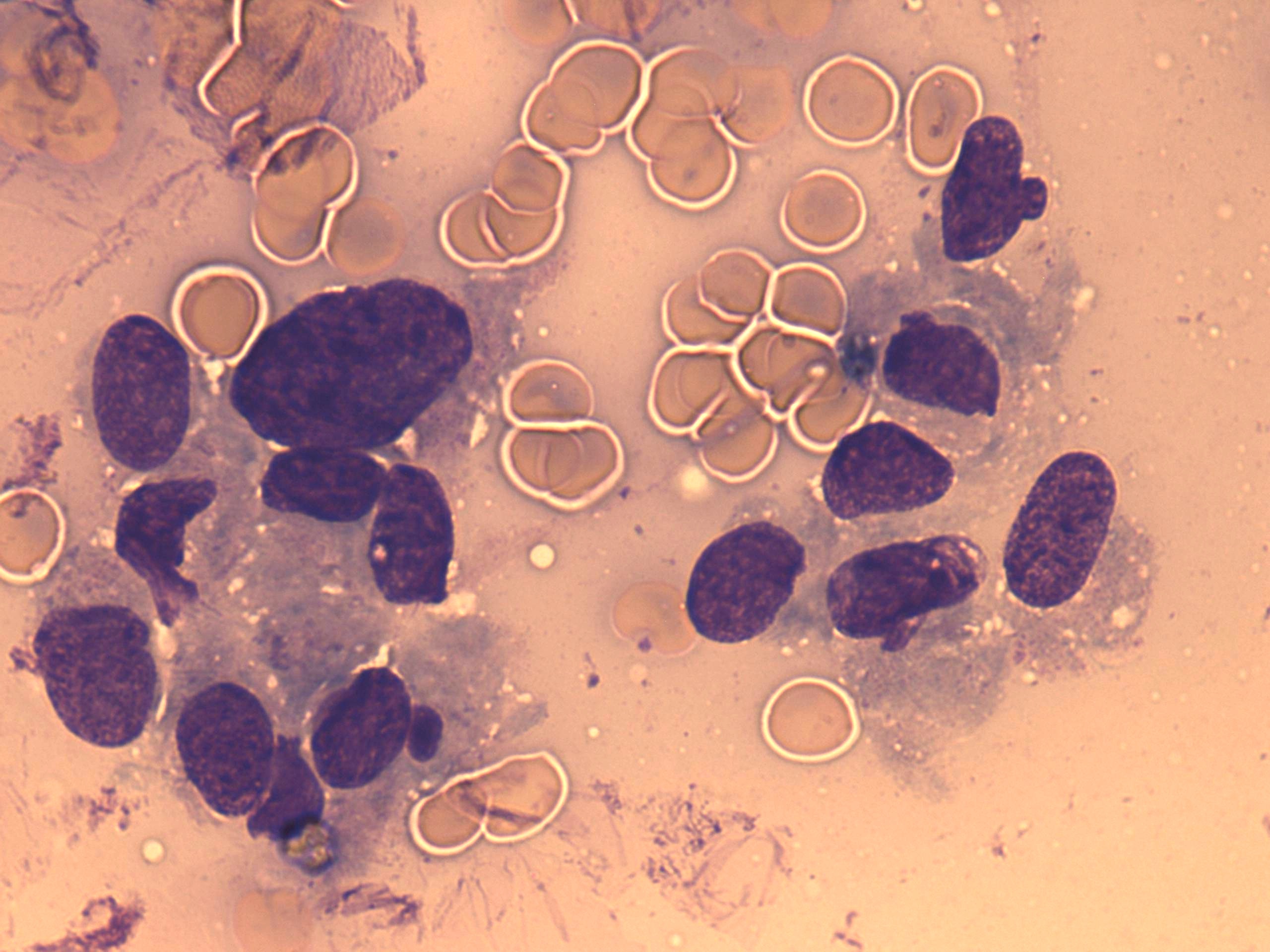
Wright-Giemsa-staining, 1000x. There is a rosette formed by tumor cells in the left part of the image. The cytoplasm contains azurophilic granulations.