
The borders of the nodule - case 430 (ultrasonographic picture 3)
|
|

|
|
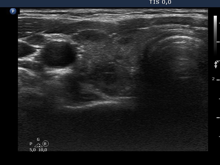
Lower part of the right lobe, transverse view. There are several discrete hypoechogenic areas. The dorsal lesion has lobulated borders.