
The borders of the nodule - case 808 (ultrasonographic picture 1)

|
|
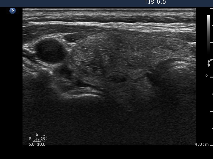
Right lobe, transverse scan. There are several discrete lesions. The lateral one presents both echogenic lines and granules, which are related to ventral cystic areas. These are back wall cystic figures caused by posterior enhancement.