
The borders of the nodule - case conp 004 (ultrasonographic picture 4)
|
|

|
|
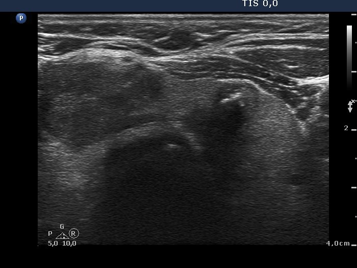
Isthmus and the left lobe, transverse scan. There is a small hypoechogenic nodule in the medial part of the left lobe. This lesion presents micro- and coarse calcifications.