
The borders of the nodule - case conp 030 (ultrasonographic picture 2)
|
|

|
|
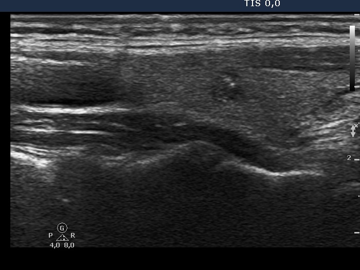
Right lobe, longitudinal view. There is a hypoechogenic nodule, which has microcalcifications and presents longer-than-wide shape.