
The borders of the nodule - case conp 037 (ultrasonographic picture 4)
|
|

|
|
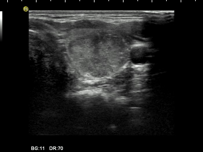
Left lobe, transverse view. There is a minimally hypoechogenic nodule with several bright hyperechogenic granules. Note the irregularity of the ventral surface.