The borders of the nodule - case conp 051 (ultrasonographic picture 5)
|
|

|
|
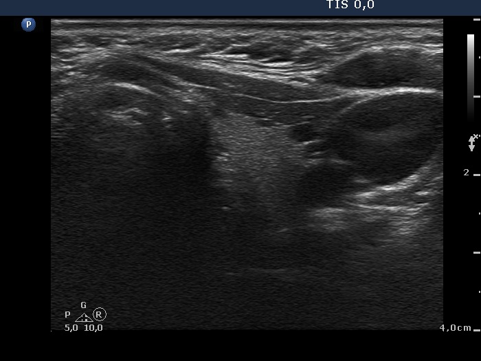
Left lobe, transverse scan. Tiny hypoechogenic foci are demonstrated in the ventrolateral part of the lobe.

2022-23 Advanced
Papillon Course
Nodule' borders
|
|
|
|
Left lobe, transverse scan. Tiny hypoechogenic foci are demonstrated in the ventrolateral part of the lobe.