
The composition of the nodule - case 1033
Examination 7 years later (ultrasonographic picture 3)
|
|

|
|
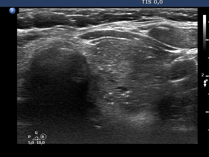
Middle part of the left lobe, transverse scan. The echogenic granules and lines are related to ventral cystic areas. Therefore, they belong very likely to figures caused by posterior back wall enhancement.