
The composition of the nodule - case 1792 (ultrasonographic picture 3)
|
|

|
|
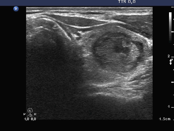
Upper part of the left lobe, another transverse view. There are bright hyperechogenic foci corresponding to non-specific granulations and back wall cystic figures. Note that the solid part is minimally hypoechoic.