
The composition of the nodule - case 2104 (ultrasonographic picture 4)
Five months after the first examination
|
|

|
|
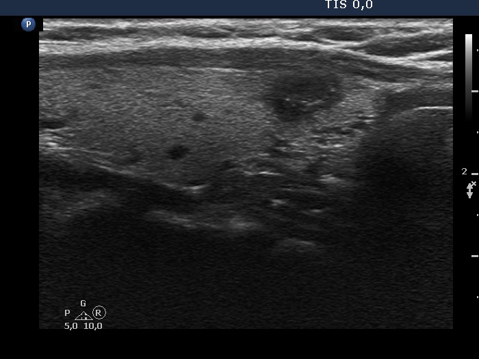
Left lobe, another longitudinal scan. The degree of blur does not reach 50%. The intranodular echogenic figures are not clearly related to ventral cystic areas. However, considering the previous examination, these can be classified as back wall cystic figures.