
The composition of the nodule - case 2176 (ultrasonographic picture 8)
|
|

|
|
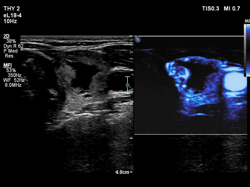
Left lobe, transverse scan, microflow imaging - after aspirating 3.5 mL brown fluid. The vessels are more easy to identify within the nodule compared with the previous situation.