
The composition of the nodule - case 6 (ultrasonographic picture 10)
|
|

|
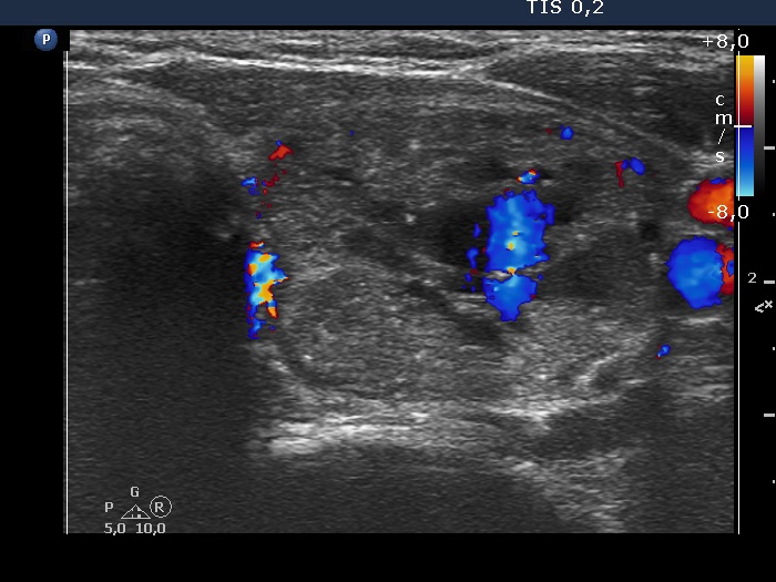
Left lobe, transverse scan, color Doppler mode - - after the removal of 8 ml bloody cystic fluid. The central blue area corresponds to the blood just refilling the cystic chambers.