
The composition of the nodule - case 653 (ultrasonographic picture 4b)
|
|

|
|
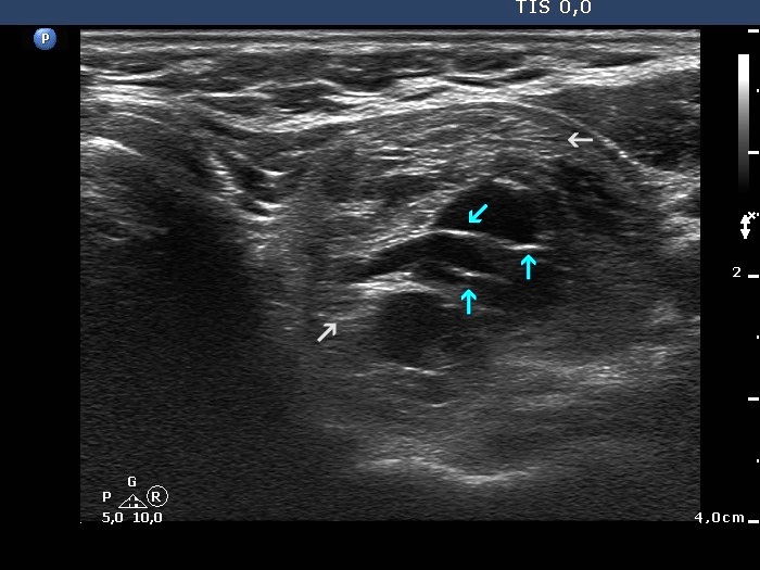
Upper part of the left lobe, transverse scan. Blue arrows point to back wall figures, lines and granules, caused by posterior enhancement. The nodule has non-specific pale echogenic figures, as well (grey arrow).