
Intranodular hyperechogenic figures - case 1423 (ultrasonographic picture 3)
|
|

|
|
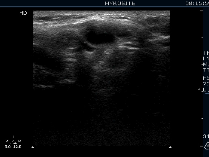
Right lobe, longitudinal view. Upper (left in the image) the granulation while lower (right in the image) the cystic lesion.