
Intranodular hyperechogenic figures - case 1425 (ultrasonographic picture 7)
|
|

|
|
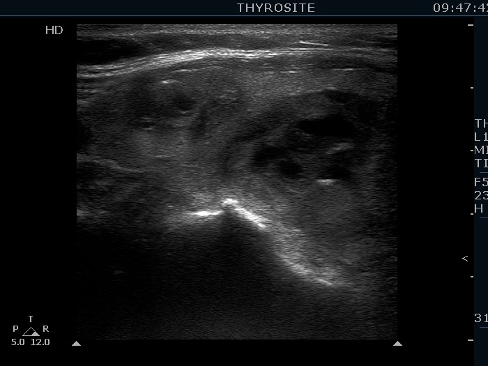
Upper part of the right lobe, longitudinal view. There is one relatively large bright hyperechogenic granule in the ventral part of the upper nodule. This might be a microcalcification.