
Intranodular hyperechogenic figures - case 1473 (ultrasonographic picture 3)
|
|

|
|
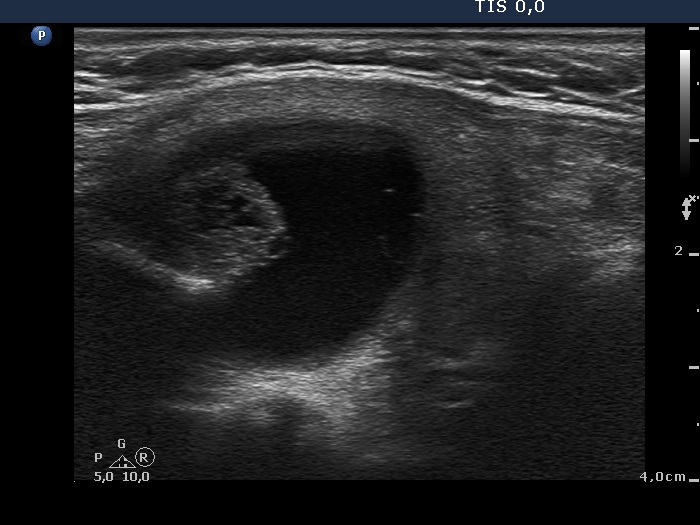
Right lobe, longitudinal view. The angle between the solid part and the wall of the nodule is acute. This image reveals that the solid part contains not only hyperechogenic granules but lines, as well. Therefore these figures are presentations of connective tissue and back wall figures depending on the lack and presence of ventral cystic areas, respectively.