
Intranodular hyperechogenic figures - case 1636 (ultrasonographic picture 6)

|

|

|
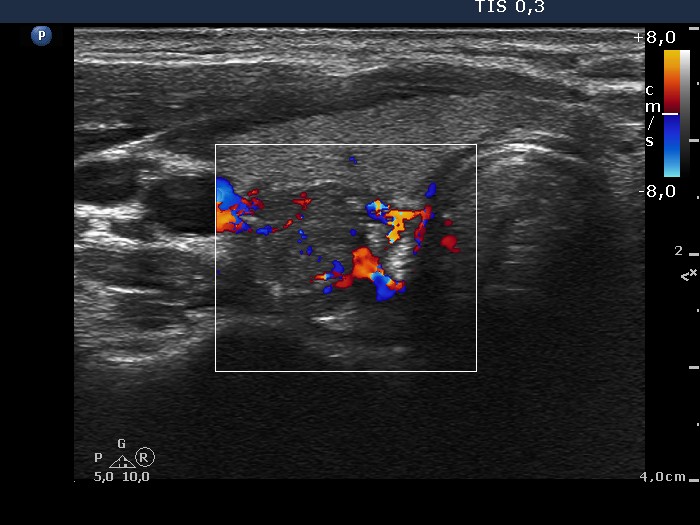
Lower part of the right lobe, transverse scan, color Doppler mode. This nodule presents irrregularly increased intranodular vascular pattern.


