
Intranodular hyperechogenic figures - case 372 (ultrasonographic picture 2)

|

|

|
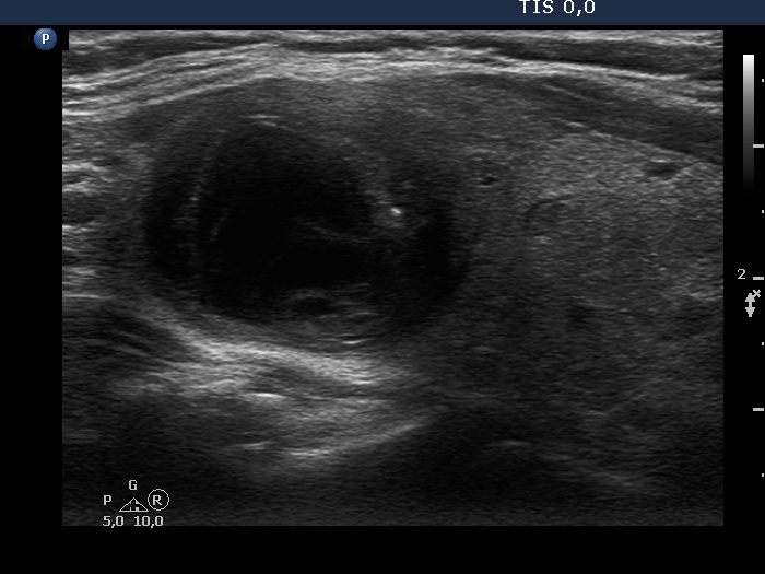
Right lobe, longitudinal view. Beside several non-specific hyperechogenic granules, there are more bright punctate echogenic foci in the solid part of the nodule. The upper (left in the image) belongs to the non-specific category while the lower two (right in the image) next to each other might be microcalcifications.


