
Intranodular hyperechogenic figures - case 386
Three weeks after the intital examination (ultrasonographic picture 1)
|
|

|
|
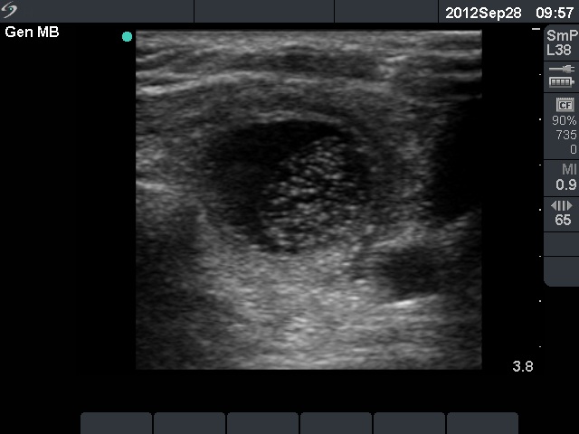
Left lobe, transverse scan. The nodule became enlarged and cystic. The borders of the nodule are ill-defined.