
Intranodular hyperechogenic figures - case 420 (ultrasonographic picture 4)

|

|

|
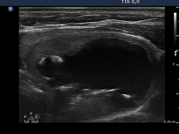
Left lobe, transverse scan. There is a central-type cyst in the left lobe. The solid part of the lesion is minimally hypoechogenic. The figures on and near to the ventral wall of the cystic area are probably aggregates of colloid crystals while the linear figures in the dorsal wall are probably caused by back wall posterior enhancement.


