
Intranodular hyperechogenic figures - case 420 (ultrasonographic picture 7)

|

|

|
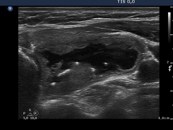
Left lobe, transverse scan - after aspirating 20 ml yellow fluid. There is a typical comet-tail artifact within the cystic fluid while the hyperechogenic figures in the dorsal wall of the cyst correspond to back wall posterior enhancements.


